


Smart Studio
Smart Studio

Lens Effect 3D
Lens Effect 3D

Optic Studio
Optic Studio
How objective Dry Eye diagnostics increase clinic revenue
April 22, 2026
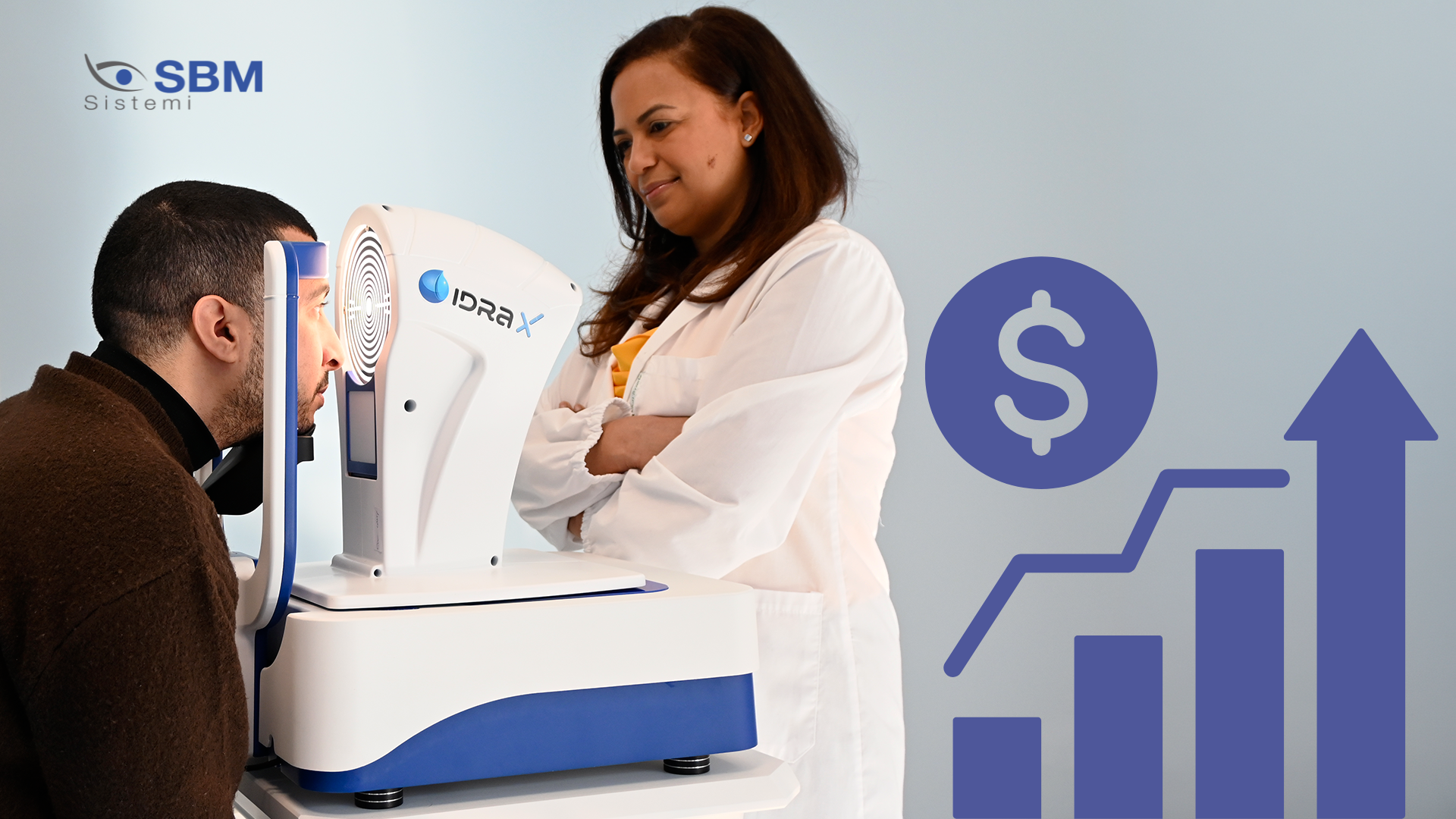
Dry eye disease is no longer a marginal complaint managed only when symptoms become severe. In modern eye care
practices, it represents both a clinical priority and a business opportunity, impacting cataract workflows,
refractive evaluations, contact lens follow-ups and long-term patient retention.
For this reason, more clinics are investing in dry eye diagnostic equipment for clinics that want to improve both
diagnostic accuracy and operational efficiency. Objective ocular surface assessment transforms dry eye from a
subjective complaint into a measurable and structured clinical pathway.
Why subjective Dry Eye management limits clinic growth
Many clinics still rely on symptoms, slit lamp observations and a limited set of traditional tests. However, dry eye
symptoms often do not correlate well with clinical signs, making diagnosis inconsistent and difficult to
standardize.
This is why the shift toward non-invasive dry eye tests is becoming increasingly important, as highlighted in SBM’s
article on best non-invasive tests for dry eye diagnosis in 2026, where reproducibility and patient comfort are
central to modern diagnostics.
From a business perspective, relying on subjective evaluation creates inefficiencies: longer chair time, unclear
communication and lower treatment acceptance. Without objective diagnostics, clinics lose both time and revenue
opportunities.
How objective diagnostics transform clinical and business performance
Objective diagnostics provide measurable data such as tear film stability, lipid layer quality and meibomian gland
structure. This makes the diagnosis clearer and more actionable, improving both clinical decisions and patient
understanding.
Technologies like a tear film analyzer device allow clinicians to move beyond subjective impressions and adopt a
structured, evidence-based approach to ocular surface evaluation.
-
Objective diagnostics improve case acceptance
When patients can see their condition, they are more likely to accept treatment. Visual tools such as meibography and tear film imaging turn an abstract problem into concrete evidence.
This is where ocular surface analysis becomes a powerful communication tool, helping clinicians explain not only the presence of dry eye but also its severity and underlying causes in a clear and visual way.
Instead of saying “you may have dry eye,” the conversation becomes data-driven and far more convincing. -
Build a complete Dry Eye treatment pathway
Revenue increases when dry eye is treated as a continuous clinical pathway rather than a one-time consultation.
A structured workflow—from screening to diagnosis, treatment and follow-up—is essential for scalability. As discussed in the article on ocular surface assessment in high-volume clinics, integrating diagnostics into daily routines improves both efficiency and consistency.
Platforms like Idra X enable this integration by combining multiple diagnostic parameters into a single workflow, while solutions such as Activa support the transition from diagnosis to in-clinic treatment. -
Reduce chair time and improve workflow efficiency
Contrary to common belief, objective diagnostics often reduce chair time rather than increase it.
Clinics that implement structured ocular surface assessment in high-volume settings can avoid repeated tests, reduce uncertainty and streamline decision-making. This results in faster consultations and a more efficient use of clinical resources.
Standardization is the key: when data is consistent, decisions are faster. -
Increase value in pre-surgical screening
Dry eye plays a critical role in surgical outcomes, particularly in cataract and refractive procedures.
Incorporating pre-surgical ocular surface evaluation allows clinicians to identify tear film instability early, improving measurement accuracy and reducing postoperative dissatisfaction.
This approach not only improves outcomes but also protects the clinic’s reputation and reduces costly follow-up visits. -
Improve patient experience and retention
Modern patients expect fast, comfortable and understandable exams. Non-invasive diagnostics meet all these expectations.
As explained in SBM’s content on non-invasive dry eye assessment, patient-friendly testing improves compliance and makes follow-up visits easier to manage.
A better experience leads directly to higher retention and long-term patient value. -
Differentiate your clinic in a competitive market
Many clinics claim to treat dry eye, but few demonstrate a structured and technology-driven approach.
Clinics that adopt advanced diagnostic tools and follow a clear ocular surface analysis protocol can position themselves as specialized centers, increasing both patient trust and online visibility.
This differentiation is essential in a competitive ophthalmic market where patients are increasingly informed and selective.
Which diagnostic features deliver the highest ROI?
The most effective dry eye diagnostic systems combine multiple parameters into a single platform, including tear
break-up time, lipid layer analysis, meibography and blink evaluation.
Integrated solutions such as an advanced ocular surface analyzer provide greater clinical insight and support a more
efficient workflow compared to single-purpose devices.
From diagnostics to recurring revenue
Revenue growth does not come from the device alone, but from the clinical model it enables.
A structured dry eye workflow (diagnosis, education, treatment and follow-up) creates opportunities for recurring
visits and long-term care.
By combining diagnostics with treatment solutions like Activa, clinics can move toward a complete, protocol-based
approach to dry eye management.
Clinics that rely only on subjective dry eye evaluation are limiting both clinical outcomes and business growth.
Objective dry eye diagnostics improve accuracy, enhance communication, streamline workflow and increase treatment
acceptance.
For practices evaluating dry eye diagnostic equipment for clinics, the real question is no longer whether to adopt
objective diagnostics, but how much growth is being missed without them.
Read more
Objective vs subjective meibography: why quantification matters
Dry Eye in dogs and cats: beyond the Schirmer Test
The business case for Dry Eye clinics: revenue opportunities and patient retention
How corneal topography helps detect keratoconus in early stages
Automated Dry Eye diagnosis: how technology improves repeatability and workflow
Corneal Topography and Dry Eye: why both matter before cataract surgery
Tear film layers explained: lipid, aqueous and mucin
How to build a complete Dry Eye diagnostic workflow in your clinic
How to choose a Dry Eye diagnostic device for your practice
Meibomian gland dropout: early detection with advanced imaging
Objective Optical Quality Metrics in Modern Ophthalmology
Regulatory Considerations When Importing CE Certified Ophthalmic Devices
Quantitative meibomian gland analysis: from images to objective data
OEM Opportunities in Ophthalmic Diagnostic Equipment
Best non-invasive tests for Dry Eye diagnosis in 2026
Transillumination meibography: clinical applications and advantages
What is a Tear Film Analyzer Device and how does it improve Dry Eye diagnosis?
Ocular surface assessment in high-volume clinics: efficiency without compromising accuracy
Beyond dry eye: other ocular surface disorders that impact visual quality
The role of ocular surface inflammation in post-surgical patient dissatisfaction
Ocular surface evaluation in contact lens wearers: what clinicians often miss
Environmental and lifestyle factors that alter tear film stability
Tear Film health in French Bulldogs: what the latest study using OSA-Vet® reveals
Pre-surgical dry eye screening: which parameters really influence outcomes
Optimizing the ocular surface before cataract surgery to improve surgical accuracy and patient outcomes
Dry eye and cataract surgery: managing expectations through objective preoperative evaluation
Evidence-based guidelines for cataract surgery: improving outcomes through standardized clinical practice
Meibomian gland dysfunction: why early detection changes long-term patient outcomes
Meibography and MGD: from gland imaging to clinical decision making
Non-invasive tear film assessment: clinical advantages over fluorescein-based methods
Tear film analysis explained: LLT, NIBUT and tear meniscus in clinical practice
Why traditional dry eye tests are not enough: limits of Schirmer and TBUT in modern practice
Dry eye diagnosis today: why ocular surface analysis is no longer optional

Attention: The information on this page is intended exclusively for qualified healthcare professionals.


