


Smart Studio
Smart Studio

Lens Effect 3D
Lens Effect 3D

Optic Studio
Optic Studio
Ocular surface evaluation in contact lens wearers: what clinicians often miss
February 19, 2026
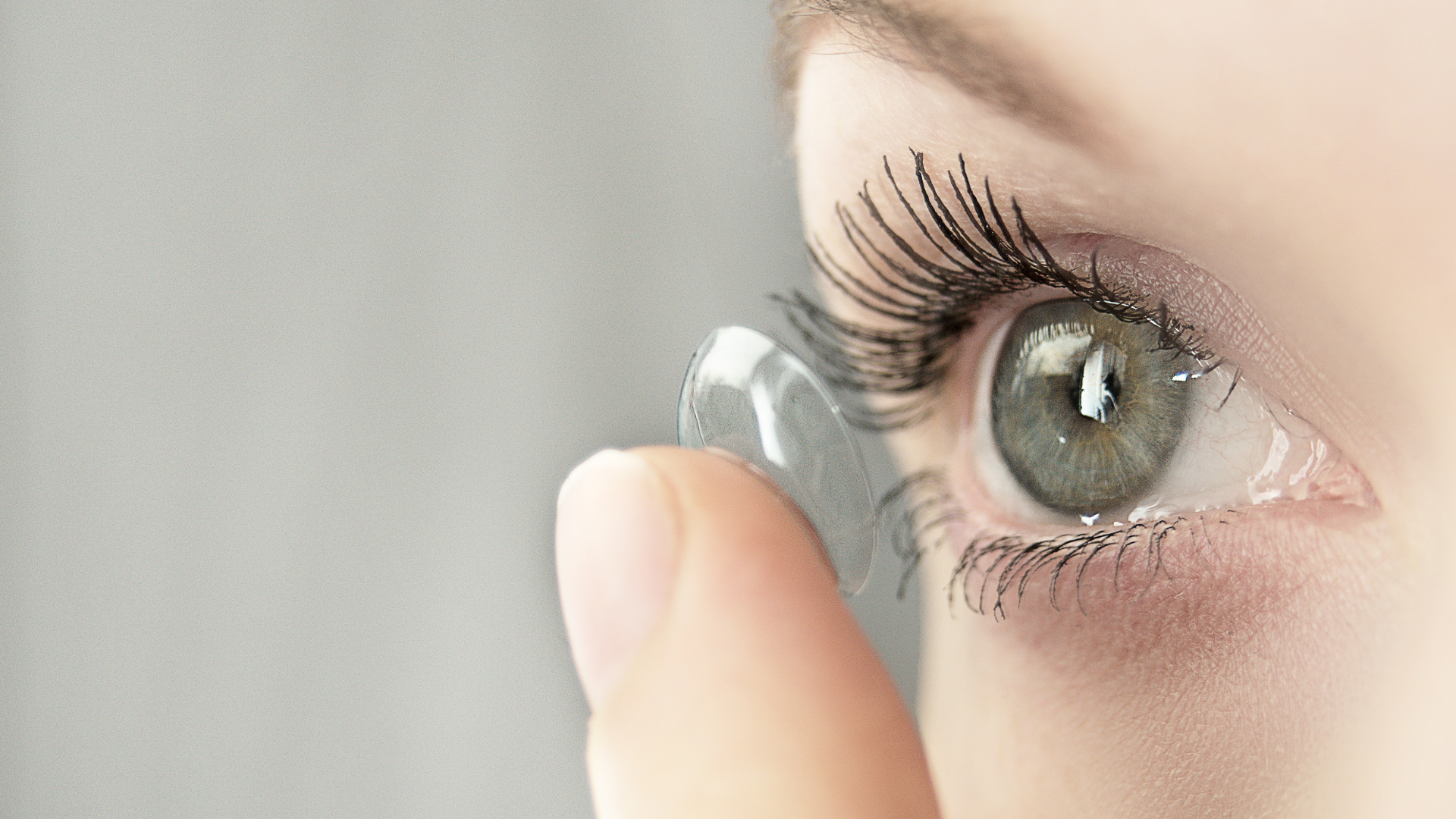
Contact lens wearers represent a unique and often underestimated subgroup in ocular surface evaluation. While many
patients report acceptable comfort and visual performance, subtle alterations of the tear film and ocular surface
may already be present long before overt symptoms develop.
Routine eye examinations frequently fail to capture these early changes, leading to underdiagnosis of tear film
instability and delayed intervention. For clinicians, recognizing what is commonly missed in contact lens wearers is
essential to preserving long-term ocular surface health and visual quality.
Contact lenses and tear film interaction
Contact lenses alter the natural tear film by dividing it into two layers: a pre-lens and a post-lens tear film.
This altered structure can:
- Increase tear evaporation
- Reduce tear film stability
- Interfere with lipid layer distribution
Even in asymptomatic patients, these changes may compromise tear film dynamics and increase susceptibility to
environmental stress.
Importantly, standard assessments performed without lenses or under ideal conditions may fail to reflect real-world
tear film behavior during lens wear.
Why symptoms are often absent or underreported
One of the most challenging aspects of ocular surface evaluation in contact lens wearers is symptom reliability.
Many patients:
- Normalize mild discomfort over time
- Attribute symptoms to lens wear rather than ocular surface changes
- Remain asymptomatic despite measurable tear film instability
As a result, clinicians may underestimate ocular surface alterations if evaluation relies primarily on symptom reporting.
Tear film instability without classic dry eye signs
Contact lens wearers may exhibit tear film instability without traditional signs of dry eye disease.
Commonly overlooked findings include:
- Reduced tear film break-up time under dynamic conditions
- Lipid layer disruption during prolonged visual tasks
- Fluctuating visual quality throughout the day
Because these patients often do not meet diagnostic criteria for dry eye disease, tear film dysfunction may go unrecognized.
Environmental and visual stress amplification
Environmental factors such as dry indoor air, air conditioning, and prolonged digital device use have a greater
impact on contact lens wearers.
Lens wear amplifies:
- Tear evaporation
- Blink irregularities during screen use
- Visual fatigue related to tear film instability
Patients may experience worsening comfort and vision later in the day, despite appearing clinically stable during morning examinations.
Limitations of standard clinical evaluation
Traditional ocular surface tests may be insufficient in contact lens wearers.
Challenges include:
- Tear film alteration caused by invasive dyes
- Measurements performed outside real-life wearing conditions
- Lack of dynamic assessment during visual tasks
As a result, clinicians may overlook early tear film dysfunction that could compromise long-term lens tolerance.
The importance of objective and non-invasive assessment
Objective, non-invasive tear film evaluation offers significant advantages in contact lens wearers.
Repeatable measurements allow clinicians to:
- Detect early tear film instability
- Monitor changes over time
- Evaluate tear film behavior without removing lenses
This approach supports more accurate decision-making, particularly when modifying lens type, wearing schedules, or advising lifestyle adjustments.
Clinical implications for long-term contact lens success
Early identification of tear film instability can:
- Improve long-term contact lens tolerance
- Reduce drop-out rates
- Prevent progression toward symptomatic ocular surface disease
By integrating tear film evaluation into routine contact lens assessments, clinicians can proactively address issues before symptoms become limiting.
Ocular surface evaluation in contact lens wearers requires a more nuanced approach than standard eye examinations.
Tear film instability, environmental sensitivity, and adaptive symptom perception often mask early ocular surface
changes. Without objective and context-aware assessment, clinicians risk overlooking factors that ultimately affect
comfort, vision, and long-term lens success.
Recognizing what is commonly missed allows for earlier intervention and more personalized contact lens management
strategies.
Read more
Objective vs subjective meibography: why quantification matters
Dry Eye in dogs and cats: beyond the Schirmer Test
The business case for Dry Eye clinics: revenue opportunities and patient retention
How corneal topography helps detect keratoconus in early stages
Automated Dry Eye diagnosis: how technology improves repeatability and workflow
Corneal Topography and Dry Eye: why both matter before cataract surgery
Tear film layers explained: lipid, aqueous and mucin
How to build a complete Dry Eye diagnostic workflow in your clinic
How to choose a Dry Eye diagnostic device for your practice
Meibomian gland dropout: early detection with advanced imaging
How objective Dry Eye diagnostics increase clinic revenue
Objective Optical Quality Metrics in Modern Ophthalmology
Regulatory Considerations When Importing CE Certified Ophthalmic Devices
Quantitative meibomian gland analysis: from images to objective data
OEM Opportunities in Ophthalmic Diagnostic Equipment
Best non-invasive tests for Dry Eye diagnosis in 2026
Transillumination meibography: clinical applications and advantages
What is a Tear Film Analyzer Device and how does it improve Dry Eye diagnosis?
Ocular surface assessment in high-volume clinics: efficiency without compromising accuracy
Beyond dry eye: other ocular surface disorders that impact visual quality
The role of ocular surface inflammation in post-surgical patient dissatisfaction
Environmental and lifestyle factors that alter tear film stability
Tear Film health in French Bulldogs: what the latest study using OSA-Vet® reveals
Pre-surgical dry eye screening: which parameters really influence outcomes
Optimizing the ocular surface before cataract surgery to improve surgical accuracy and patient outcomes
Dry eye and cataract surgery: managing expectations through objective preoperative evaluation
Evidence-based guidelines for cataract surgery: improving outcomes through standardized clinical practice
Meibomian gland dysfunction: why early detection changes long-term patient outcomes
Meibography and MGD: from gland imaging to clinical decision making
Non-invasive tear film assessment: clinical advantages over fluorescein-based methods
Tear film analysis explained: LLT, NIBUT and tear meniscus in clinical practice
Why traditional dry eye tests are not enough: limits of Schirmer and TBUT in modern practice
Dry eye diagnosis today: why ocular surface analysis is no longer optional

Attention: The information on this page is intended exclusively for qualified healthcare professionals.


