


Smart Studio
Smart Studio

Lens Effect 3D
Lens Effect 3D

Optic Studio
Optic Studio
Listen the video
Advanced Ocular Surface Analysis
for Veterinary Care
A comprehensive platform designed to assess tear film quality,
ocular surface conditions and Meibomian gland morphology in animals.
Combining advanced imaging technology with an intuitive interface,
OSA-Vet supports accurate and efficient veterinary eye examinations.
Take a closer look
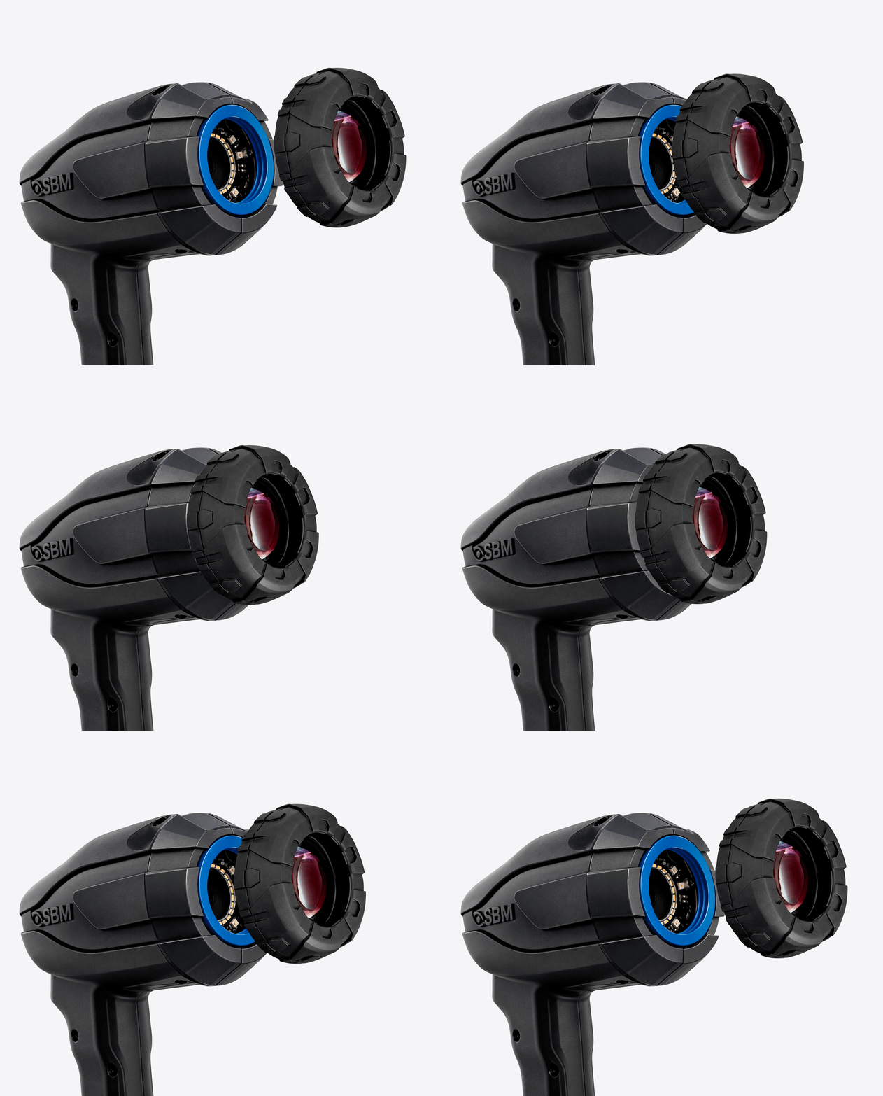
Inteferometry cone
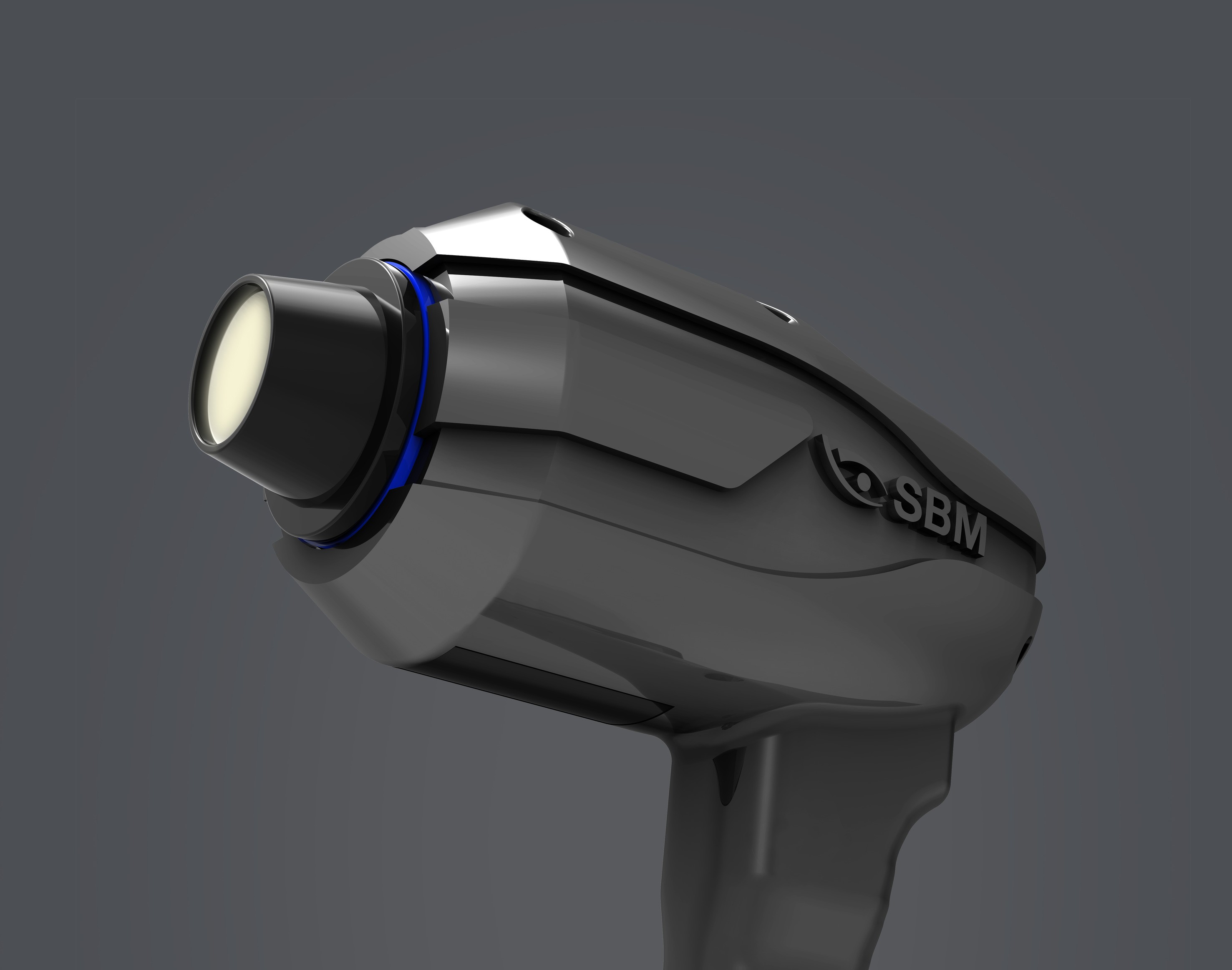
Inteferometry cone. allows visualization and assessment of the tear film's lipid layer, supporting veterinarians in evaluating tear film quality and stability.
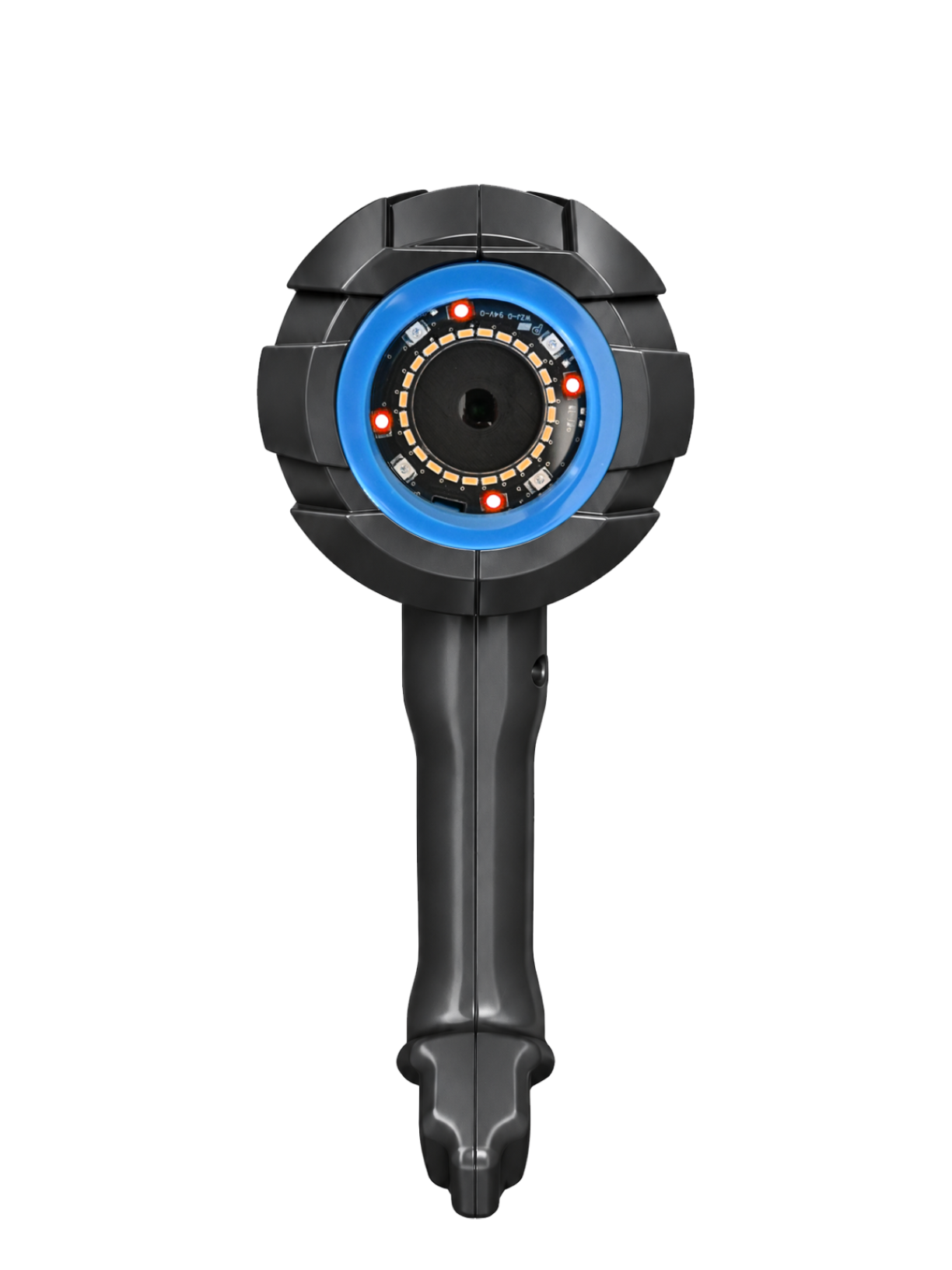
Blefarite cone
Blefarite cone. allows detailed observation of the eyelid margin and identification of signs of blepharitis, supporting veterinarians in the assessment and monitoring of eyelid abnormalities.
Infrared
Infrared. Infrared technology for comfortable, non-invasive visualization of Meibomian gland structure and potential periocular abnormalities.
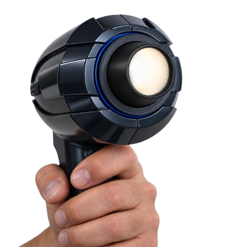
Convenience and Precision at Your Fingertips.
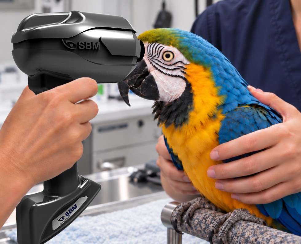
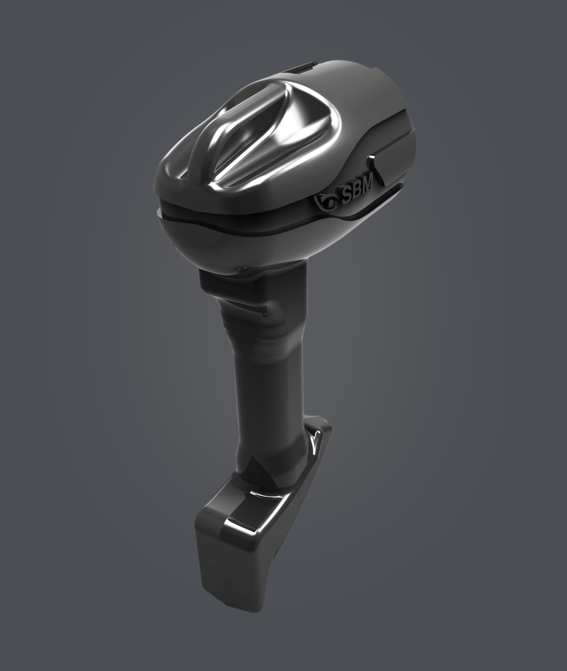
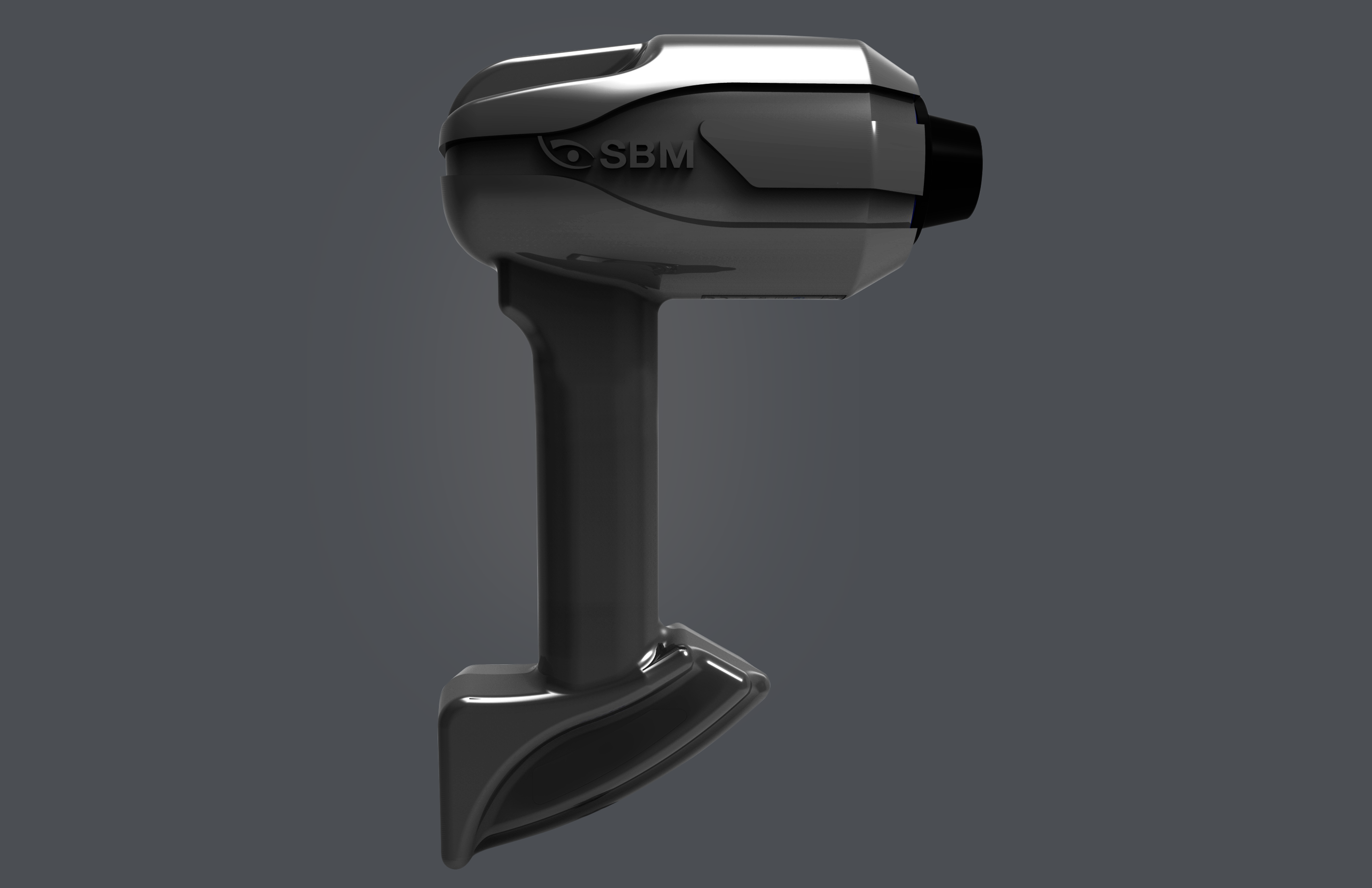
Convenience and Precision at Your Fingertips. Thanks to its ergonomic design and lightweight, easy-to-handle structure, OSA-Vet makes eye examinations in animals simple and intuitive, facilitating the veterinarian’s work while minimizing discomfort for the patient.



Explore numerous exam options

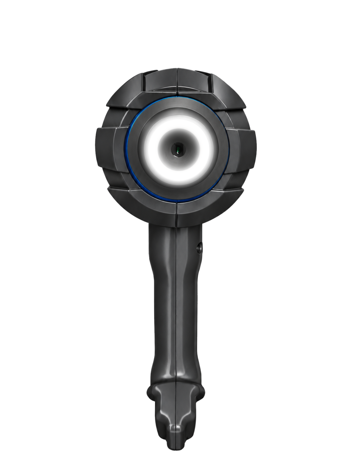


Interferometry
OSA-Vet helps diagnose Dry Eye Disease (DED) by analyzing the Precorneal Tear Film (PTF), focusing on deficiencies in the lipid, aqueous, and mucin layers. The system uses scattered light to examine the lipid layer and provides tools for immediate interpretation and follow-up, along with accessories like grids for evaluating tear film quality and structure.
Meibography
Meibography is used to assess the condition of meibomian glands, helping diagnose Meibomian Gland Dysfunction (MGD). The OSA-VET system captures images of these glands and provides tools for analysis, such as image enhancements, gland loss classification, and comparison over time. Automatic detection of gland position and measurement of gland size is also integrated into the system.
NIBUT
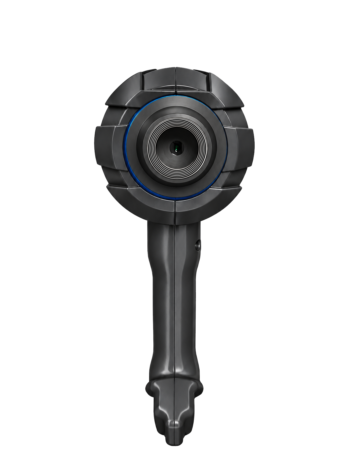
The OSA-VET enables the study of tear film stability through N.I.B.U.T., a non-invasive method that avoids the use of fluorescein, unlike the BOT test. Placido discs are also used for assessing corneal topography and tear film defects.
Tear Meniscus
The tear meniscus observed on the eyelid margins provides valuable information about tear volume, with its height, regularity, and shape being important factors. In dogs and cats, the Pre-corneal Tear Film (PTF) is about 8 µm thick on the central cornea but thicker at the meniscus, which can help assess the lacrimal glands' aqueous production.
WTW
Evaluation of corneal diameter from limbus to limbus (white-to-white distance, WTW).
One Device. Multiple Examinations.
OSA-Vet brings together a wide range of ocular surface examinations in a single integrated platform.
From tear film assessment to Meibomian gland imaging, it provides valuable information to support
veterinary professionals in evaluating dry eye and monitoring ocular surface health.
Technical Specifications

Image Resolution
5 MP

Focus
Auto, Manual

Acquisition Mode
Multi shot, video

ISO Management
Variable

Camera
Colored, sensitive to infrared (NIR), yellow-filtered

Cones
Interferometry Cone, Placid Cone

Light Source
Infrared led - blue, red and white led
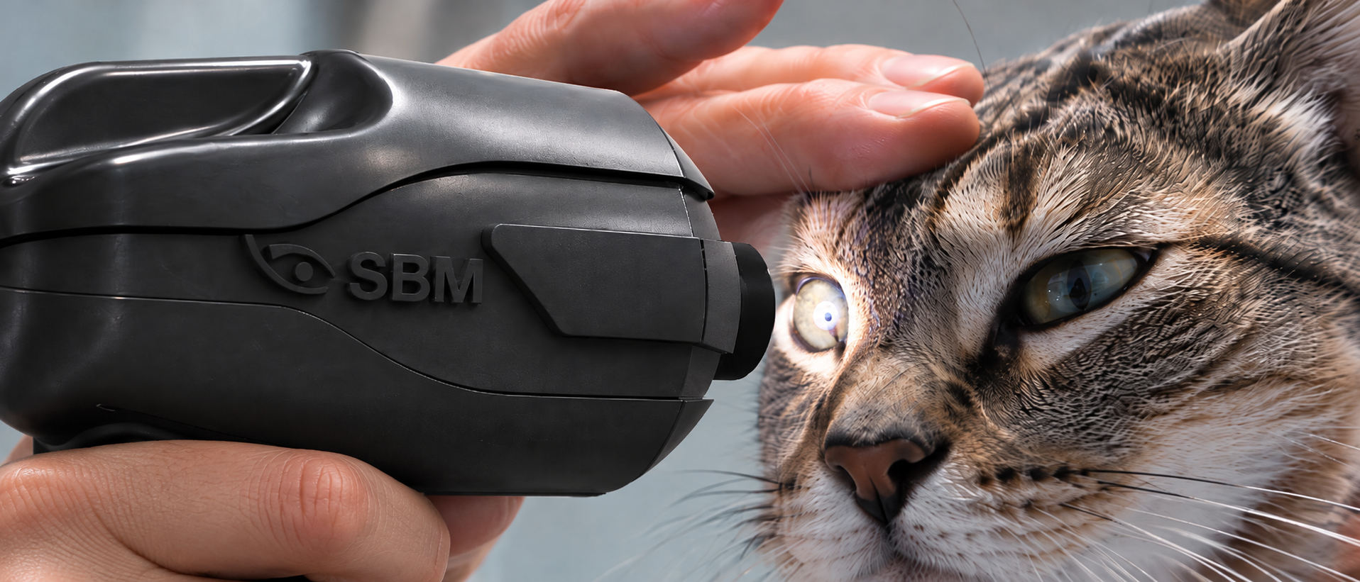
See Every Detail
With high-resolution imaging, multiple illumination options and interchangeable cones,
OSA-Vet offers versatile tools for detailed ocular surface observation.
Capture images and videos, examine corneal reflections and
document findings directly through dedicated software.
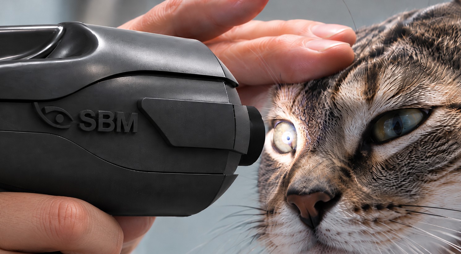
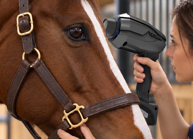
Designed for Animals. Made for Veterinarians.
Lightweight, handheld and easy to operate, OSA-Vet adapts naturally to the needs of veterinary examinations.
Its ergonomic design allows veterinarians to perform ocular surface assessments with minimal handling,
making examinations more convenient for both animals and professionals
A Closer Look at Veterinary Eye Health
Discover how OSA-Vet can enhance your veterinary ophthalmology practice with
advanced ocular surface imaging and a complete range of examinations
Many accessories,
better experience
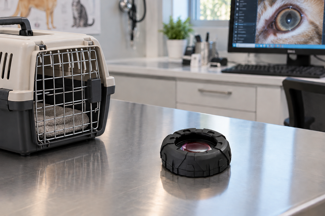
Blepharite cone
Blepharite cone
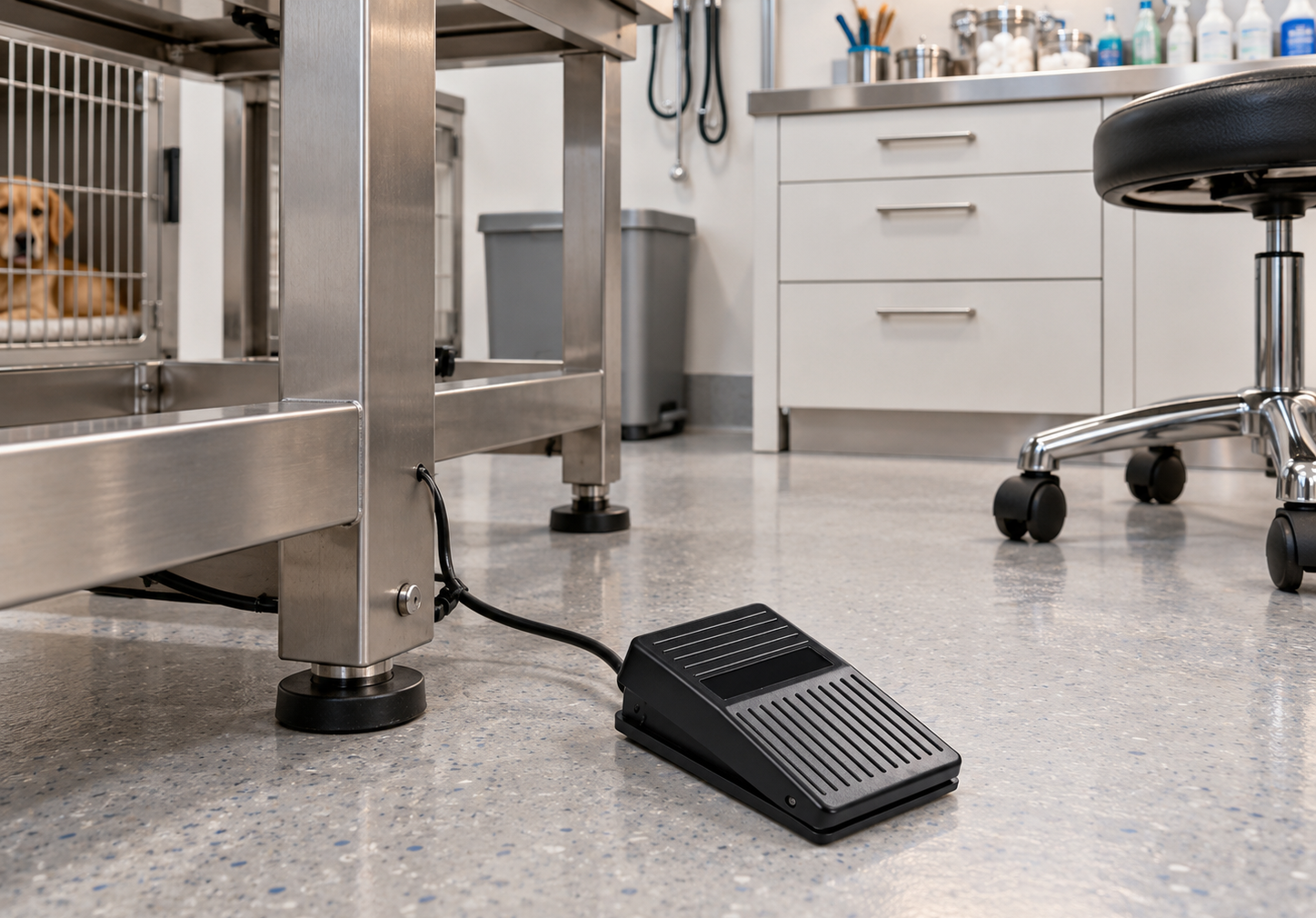
Foot pedal
Foot pedal
Mini Computer
Compact form factor computer conveniently configured and tested with SBM Software to properly run your new device with the convenience of a plug and play solution.

Extended USB Powered Cable
Dimensions 7 m; Powered USB 3.0 extension cable for camera to computer connection.

Videos





Attention: The information on this page is intended exclusively for qualified healthcare professionals.


