


Smart Studio
Smart Studio

Lens Effect 3D
Lens Effect 3D

Optic Studio
Optic Studio
Take a closer look
Inteferometry panel
Inteferometry panel. Light scattering technology enabling advanced tear film analysis for precise assessment of the lipid layer thickness.
Placido light
Placido light. High-precision Placido disc illumination system for clear and reliable corneal surface acquisition and non-invasive tear film stability evaluation.
Infrared
Infrared. Infrared technology for comfortable, non-invasive visualization of Meibomian gland structure and potential periocular abnormalities (Demodex).
Automatic movement
Automatic movement. Fully automated movement system ensuring fast, repeatable, and operator-independent examinations.
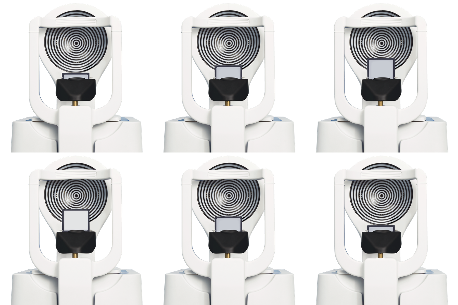
Transillumination everter
Transillumination everter. Integrated transillumination system that enhances detailed visualization of the Meibomian glands, supporting more accurate diagnosis.



Explore numerous exam options
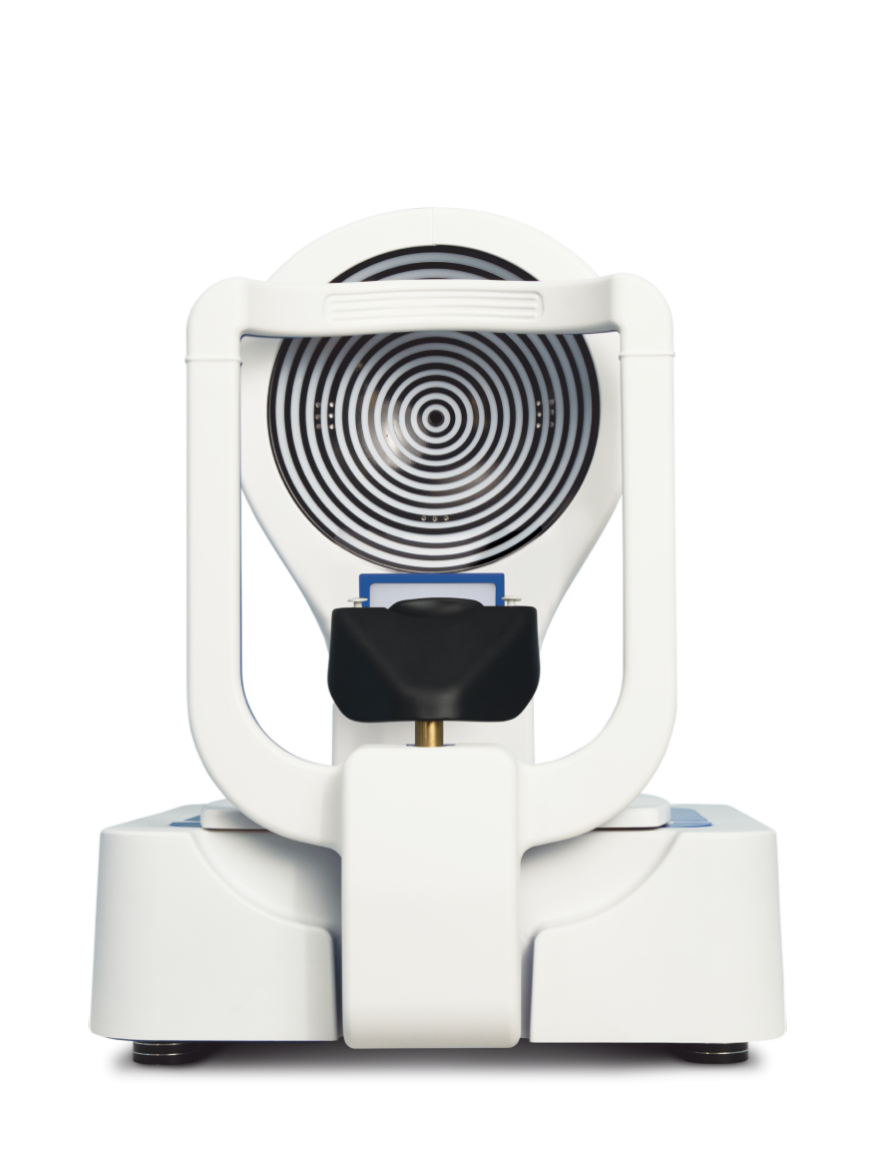
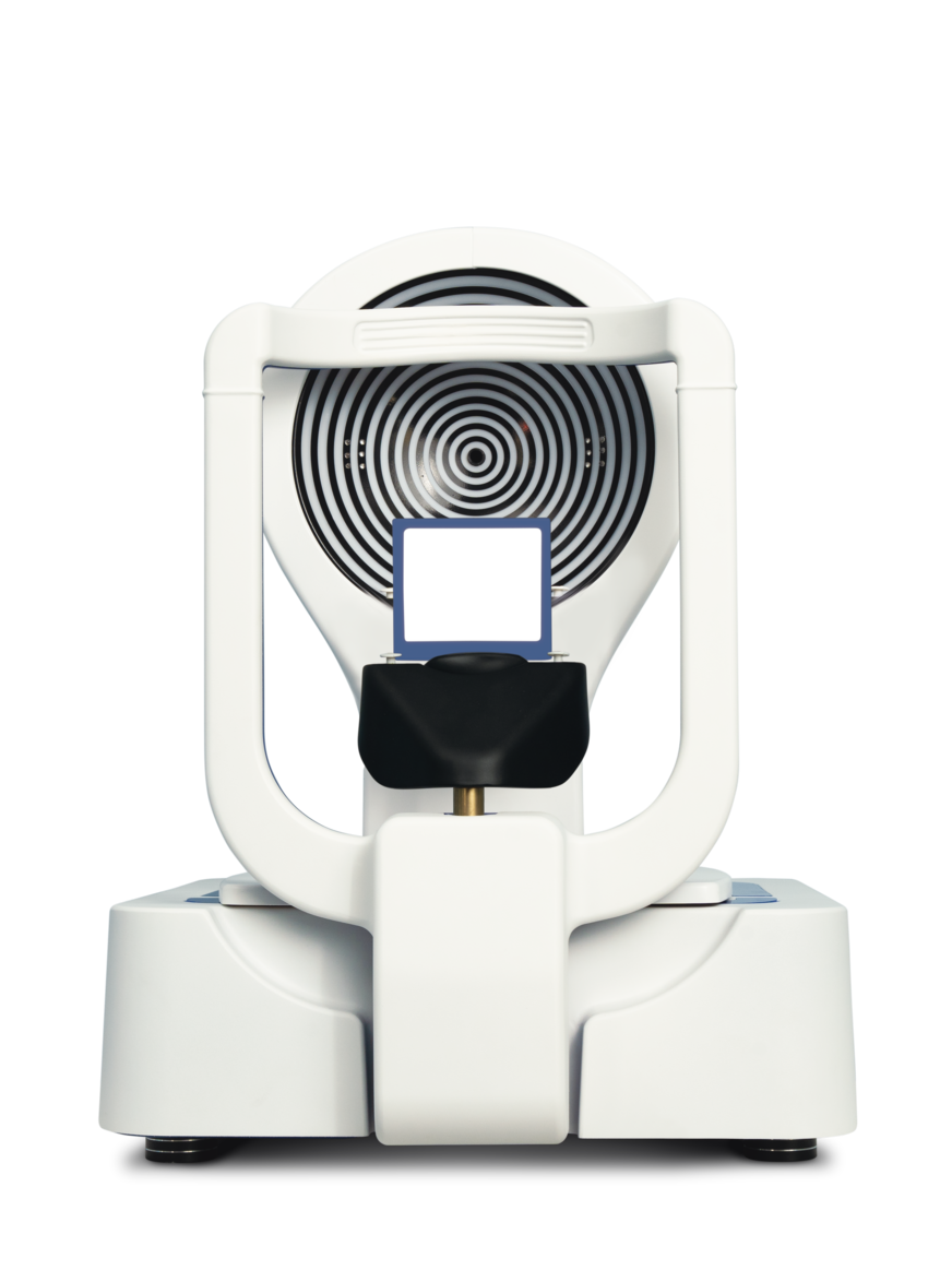
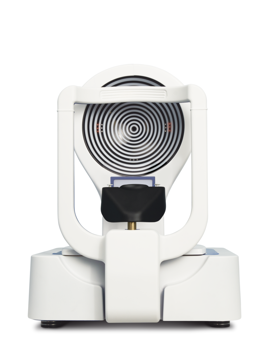
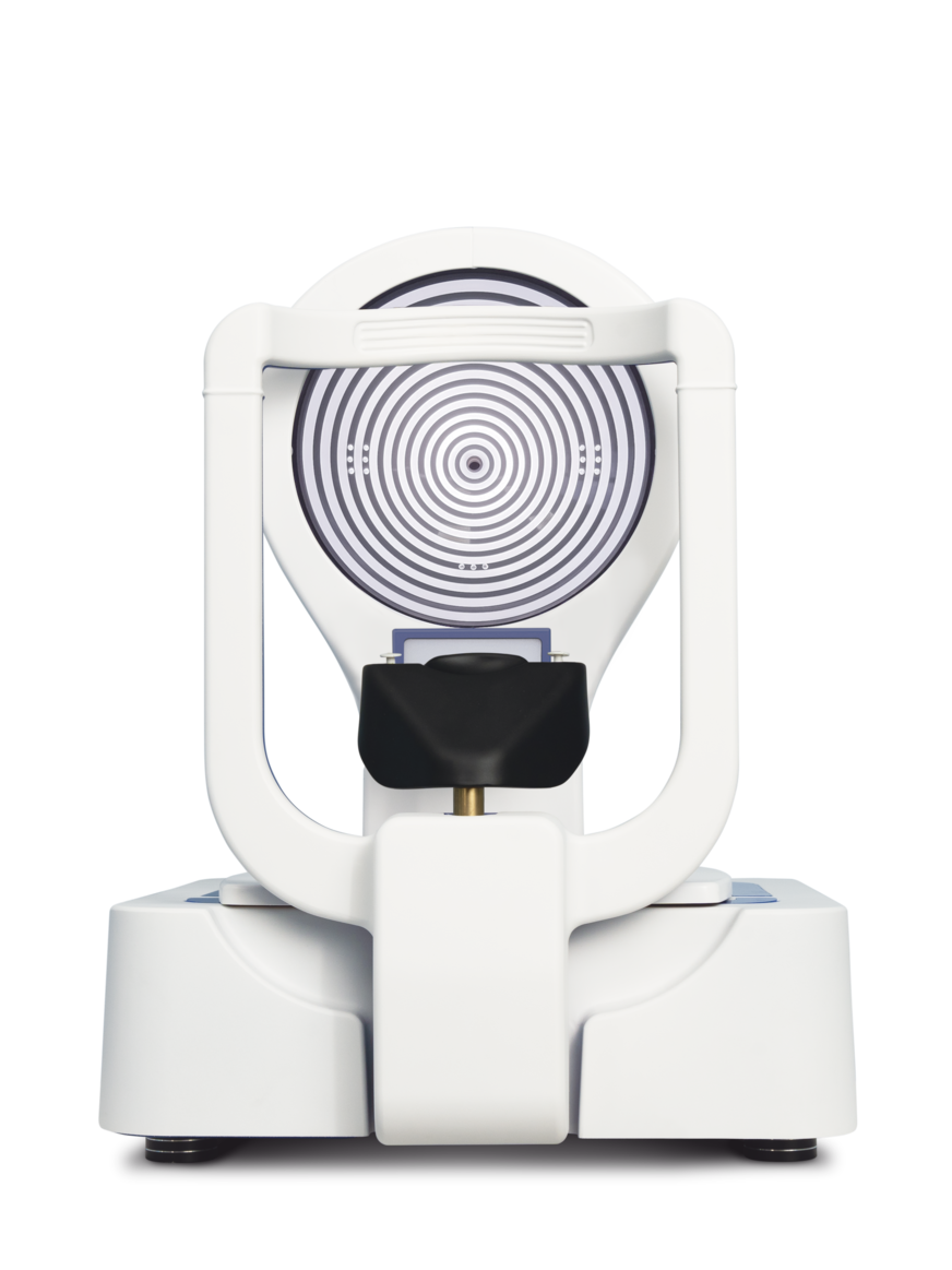

Topography
Corneal topography provides a detailed mapping of corneal curvature, supporting the evaluation of corneal shape and visual performance. It plays a key role in keratoconus screening, refractive procedures, and contact lens fitting by delivering precise and comprehensive surface data.
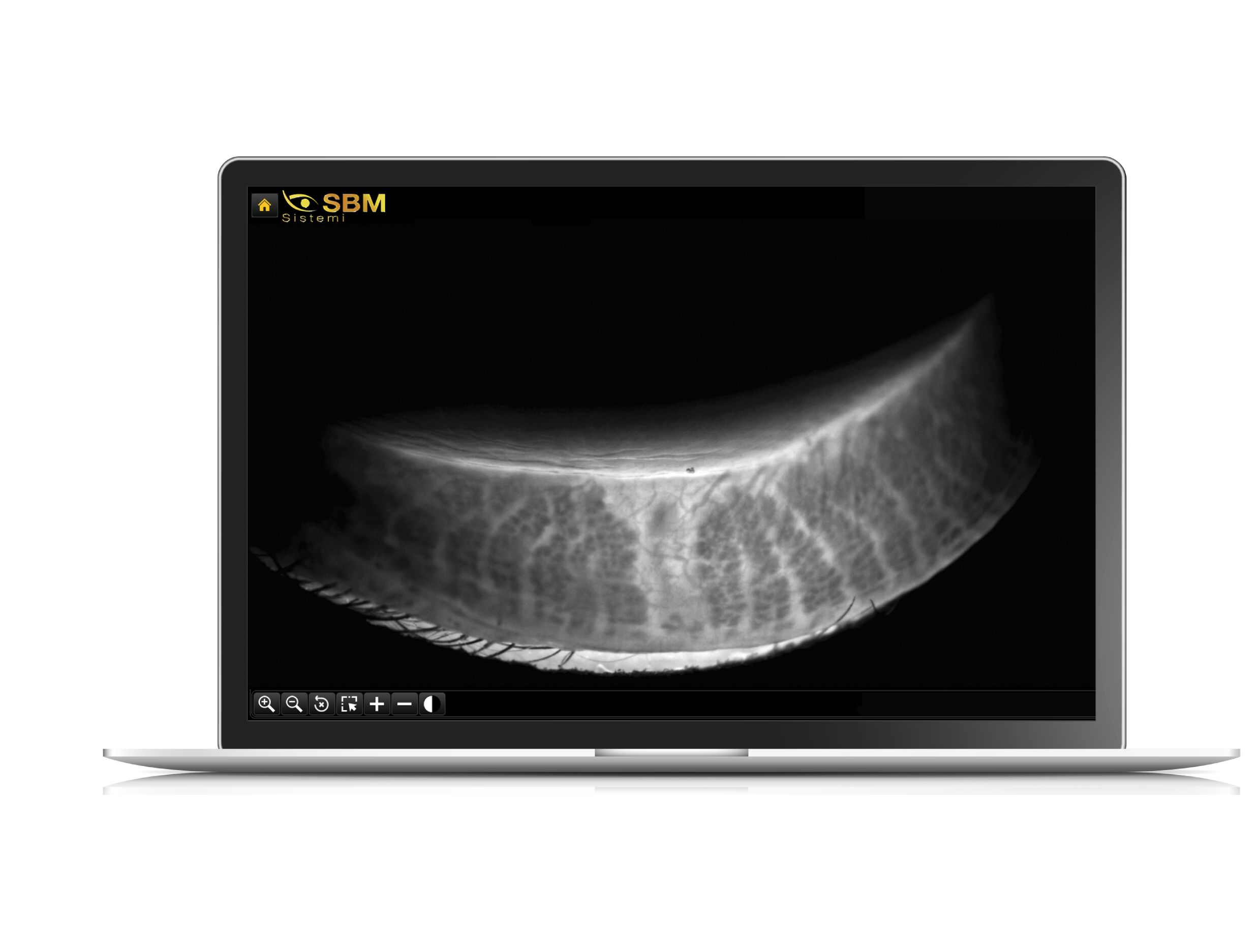
Transilluminated Meibography
Transillumination meibography enhances gland visualization through infrared light, revealing subtle structural changes and areas of dysfunction. This technique improves diagnostic depth and supports clinical interpretation.
3D Meibography
3D Meibography introduces volumetric visualization of the Meibomian glands, enabling deeper analysis of their morphology. This approach enhances the understanding of gland alterations and supports more informed clinical evaluation and patient communication.
Auto Interferometry
Auto-interferometry performs automated analysis of the tear film lipid layer, transforming a traditionally qualitative test into a standardized and objective assessment. It helps identify evaporative components of dry eye with high reproducibility.
Auto Tear Meniscus
Automatic tear meniscus evaluation measures tear volume with speed and consistency. The system ensures reliable data acquisition, supporting the identification of aqueous deficiencies and improving diagnostic confidence.
Blepharitis
The device enables accurate observation of eyelid margin conditions, facilitating the detection of blepharitis and associated signs such as debris or inflammation. This supports targeted evaluation and follow-up of eyelid disorders.
Bulbar Redness
Bulbar redness analysis provides a structured assessment of ocular surface inflammation. Captured images can be compared using standardized grading scales, allowing objective monitoring of disease progression over time.
Eye Blink
Blink analysis evaluates eyelid dynamics and blink completeness, offering insights into tear film distribution and gland function. This information helps identify behavioral or functional factors influencing ocular surface stability.
Interferometry
Interferometry enables detailed examination of the tear film lipid layer, supporting the identification of instability and evaporative mechanisms. It provides essential data for understanding dry eye etiology and guiding treatment.
Meibography
Meibography captures high-definition images of the Meibomian glands using infrared technology. It allows clinicians to assess gland integrity, structure, and potential dysfunction with clarity and precision.
NIBUT
NIBUT measures tear film stability by analyzing the time before break-up occurs after blinking. This non-invasive assessment provides reliable parameters for evaluating ocular surface health and monitoring changes over time.
Pupillometry
Pupillometry measures pupil size and responsiveness across different lighting conditions. It supports advanced diagnostics, including refractive planning and contact lens fitting, through accurate and dynamic evaluation.
Tear Meniscus
Tear meniscus assessment provides insight into the quantity of the aqueous component of the tear film. It plays a key role in distinguishing different forms of dry eye and guiding therapeutic strategies.
Vigo Suggestions
The integrated algorithm generates personalized treatment suggestions based on collected diagnostic data. This feature supports clinicians by simplifying decision-making and enabling a structured therapeutic approach.
WTW
White-to-white measurement determines the horizontal corneal diameter with precision. This parameter is essential for contact lens fitting and contributes to accurate anterior segment evaluation.
Videos
Discover the perfect solution for you
Exams
os1000x plus
os1000x
Transillumination Meibography
X
X
Automatic eye detection
X
X
Automatic focus
X
X
Automatic acquisition
X
X
Topography Map
X
X
Comparison Map
X
X
Axial Map
X
X
Tangential Map
X
X
Refraction Map
X
X
Differential Map
X
X
Corneal height Map
X
X
Pupillometry
X
X
White to white measurement
X
X
Keratoconus screening
X
X
Contact lens fitting simulation
X
X
NIBUT
X
X
Meibography
X
X
3D Meibography
X
X
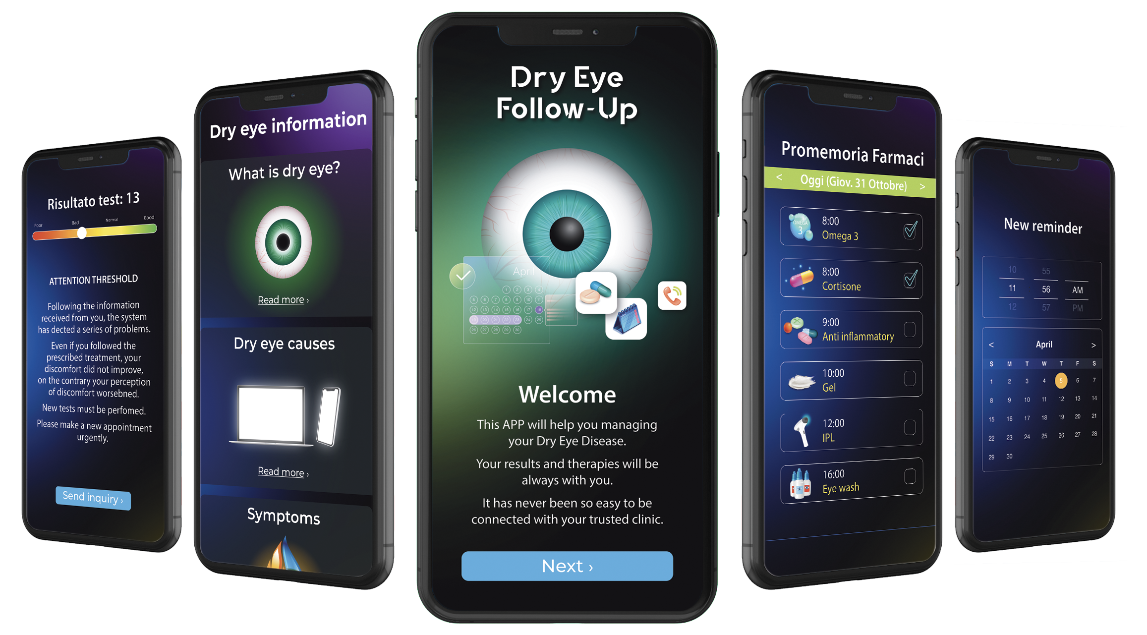
Smartphone App "Dry Eye Follow-Up"
X
X
Wizard procedure
X
X
Treatment protocol section
X
X
Interferometry
X
Auto
Tear Meniscus
X
Auto
Blink Quality
-
X
Blepharitis
-
X
Ocular redness classification
X
X
The differences between the two versions
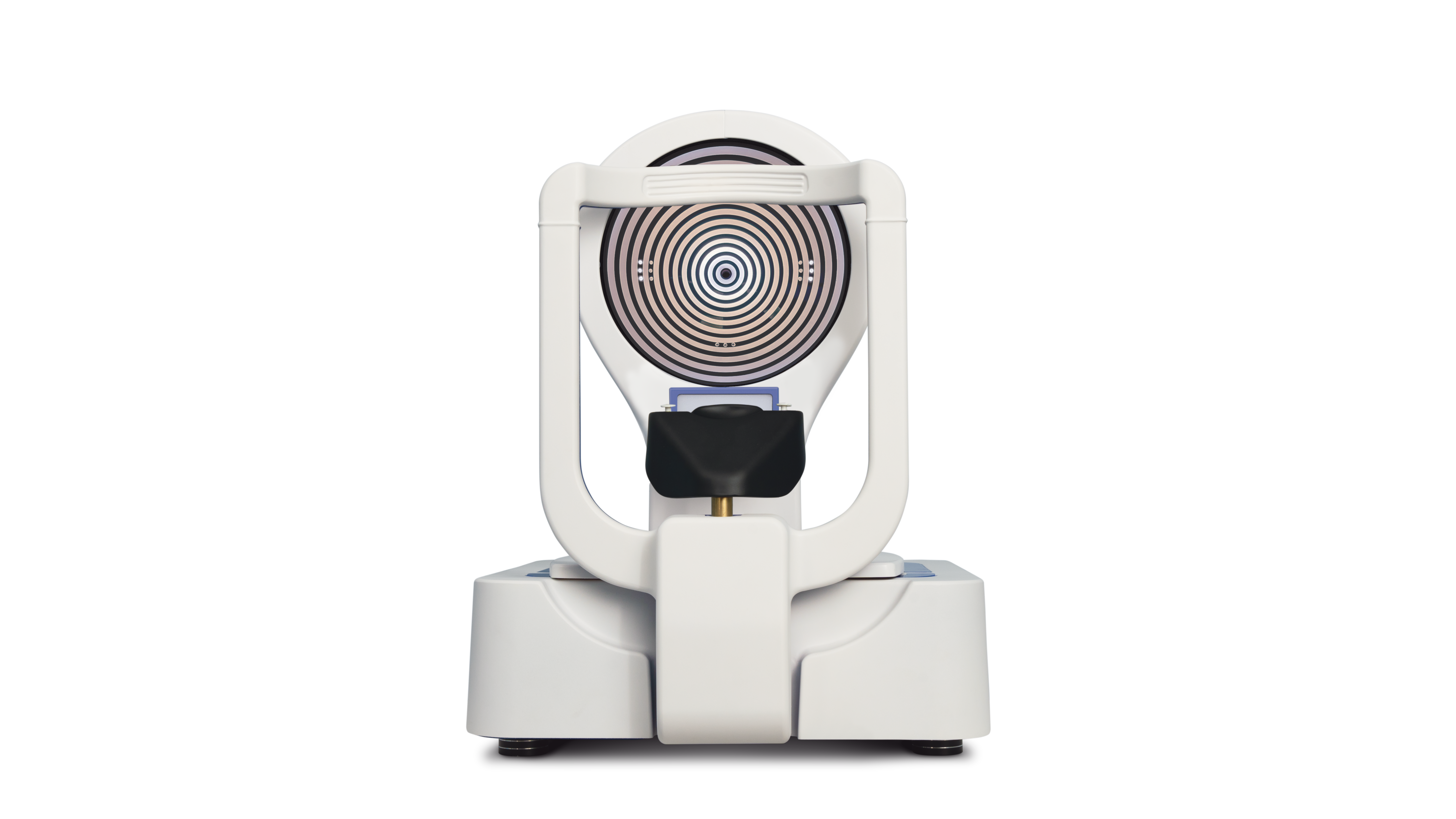
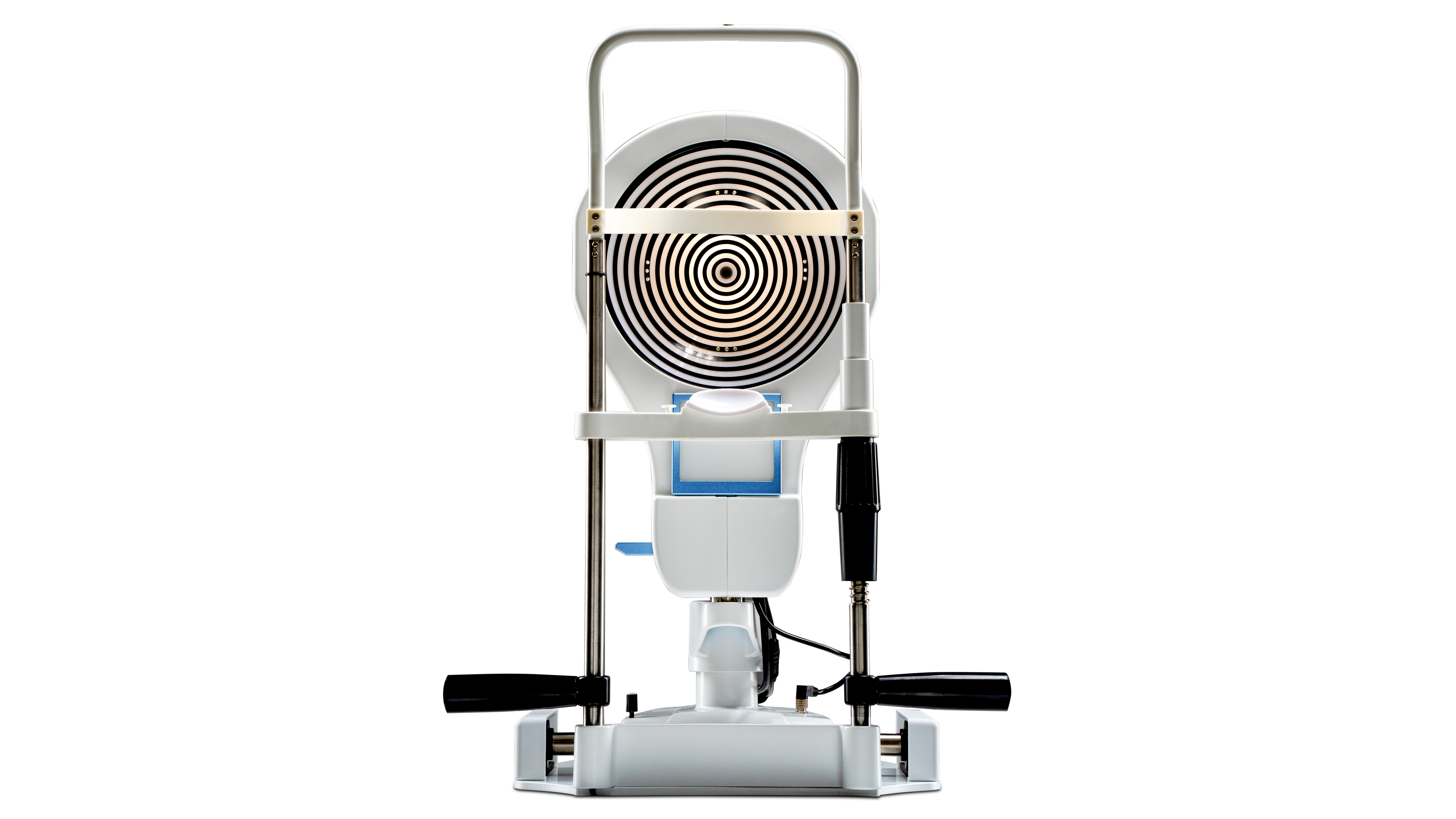
Many accessories,
better experience
Transillumination everter
Transillumination everter

Mini Computer
Compact form factor computer conveniently configured and tested with SBM Software to properly run your new device with the convenience of a plug and play solution.

Opthalmic Table
Curved table top (V_shape) with aluminum lifting column, for 2 devices. Table top size 1040mm x 550mm.

Device Cover
Cover for devices.

Extended USB Powered Cable
Dimensions 7 m; Powered USB 3.0 extension cable for camera to computer connection.

Technical Specifications

Rings
24

Measured points
8640

Camera resolution
8 Megapixel

Photo resolution
2592x1944 JPEG format
Upscaled analyzed image resolution
23 Megapixel

Acquisition mode
Single shot, multishot, video

Focus
Manual and automatic focus

ISO management
Variable

Image Color
Colours - Infrared (IR)

Lighting source
Infrared led - White led - Blue led

Working distance
60mm - 90mm from the center of the placid

Output 1
USB 3.0

Electromagnetic compatibility (EMC)
IEC 60601-1-2 (2015)

Supply voltage
100-240 V

Device operating voltage
24V - 5V - 3,3V

Dimensions
40cm (L) x 52–55cm (H) x 47cm (D)

Weight
12 Kg

Accuracy
Class A according to UNI EN ISO 19980-2021

Corneal Coverage
10mm
Product innovations
Eye tracking technology
Autofocus
Voice guidance
Integrated base and chinrest
Transillumination examination
Together, they perform real-time acquisition, analysis, and automatic evaluation of all topographic and ocular surface parameters.
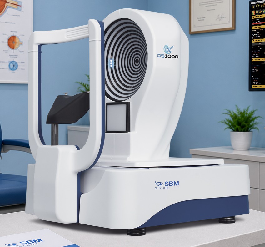
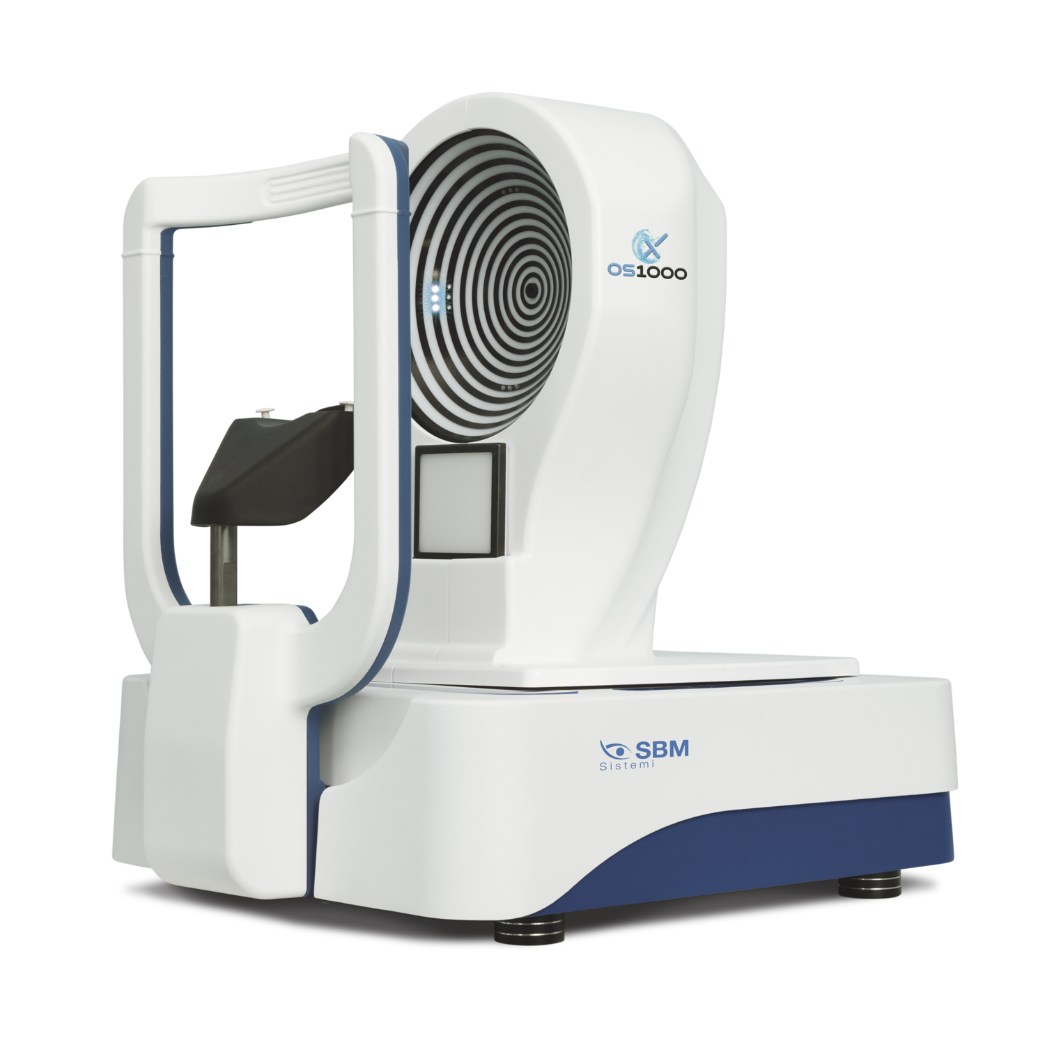
OS1000 X is the ultimate expression of
innovation in ocular surface analysis
An accurate and comprehensive assessment of the ocular surface is essential for early
diagnosis, therapeutic planning, and long-term management of its conditions.
The integrated OS1000 X system improves diagnostic accuracy, supporting more
informed therapeutic decisions and better results for patients
OS1000 X allows for the objective evaluation of:
ANTERIOR CORNEAL ABERRATIONS
CORNEAL TOPOGRAPHY
AUTOMATIC CLASSIFICATION OF DRY EYE SUBTYPES
EYELID MARGIN ABNORMALITIES
Examinations are performed through an automated workflow, simplifying both data acquisition and analysis.
Improved resolution for the analyzed image
An AI-based algorithm increases the resolution of the captured image up to 23 megapixels.
The ultra-high-quality image is then processed, achieving new levels of precision in
corneal topographic measurement based on Placido rings

Standard corneal
topographers

OS1000 X
by SBM Sistemi
Key features and workflow
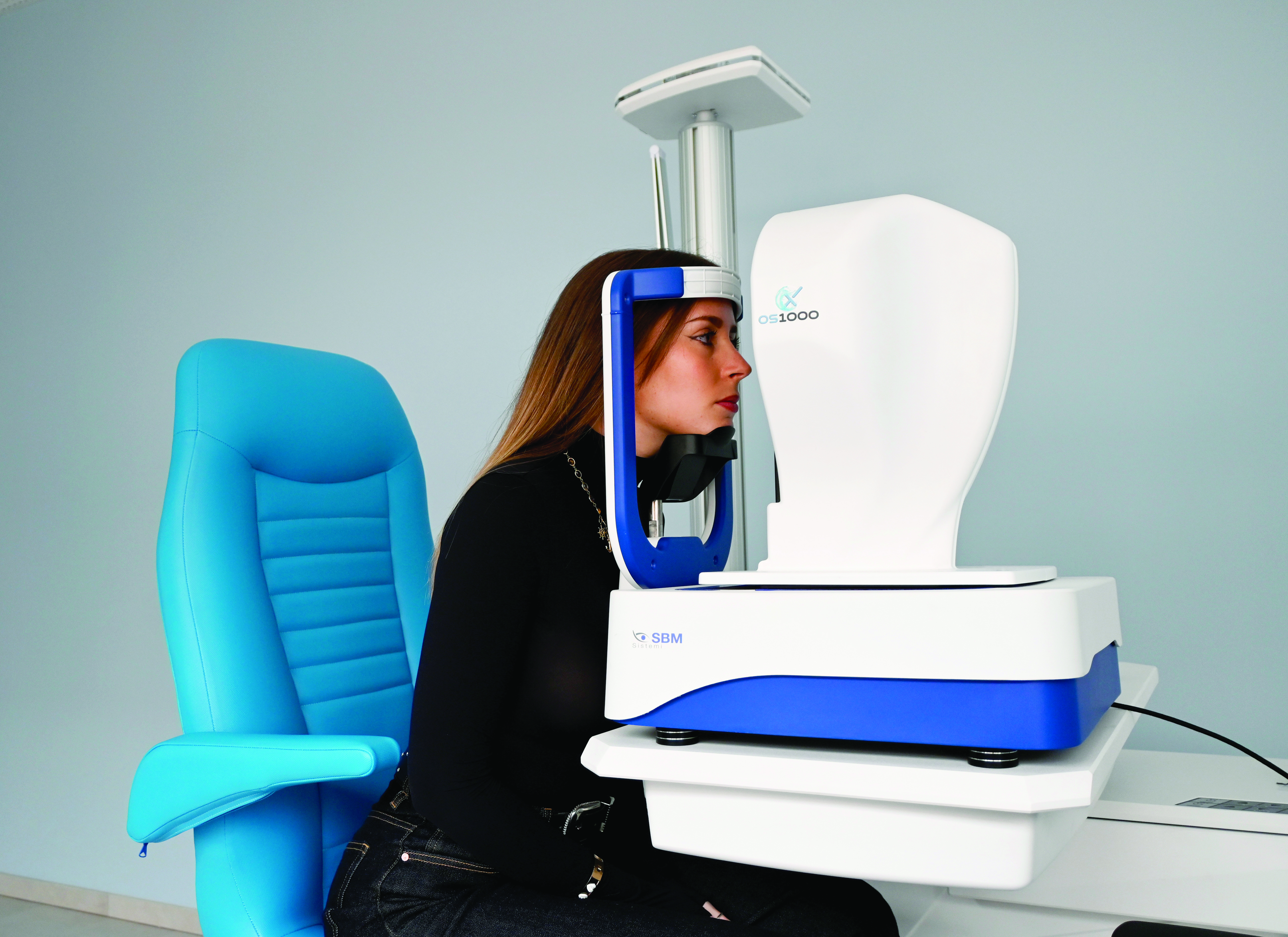
Equipped with an automated base, the OS1000 X initiates the data acquisition process with a simple click.
Once activated, the device aligns, focuses, and automatically captures the required images, guided by an intuitive software interface.
A guided procedure coordinates the process, automatically performing the selected diagnostic
tests and processing the results with minimal user intervention.
Why choose the OS1000 X?
Disorders of the ocular surface, particularly Dry Eye, are very common and require reliable diagnostic tools for an accurate
patient assessment. Severe Dry Eye can, in fact, affect the health and shape of the cornea, also impacting topographic results.
Modern lifestyles, characterized by prolonged screen use, air conditioning, and pollution, promote the chronic development of this condition.
Implementing an automated, structured, and personalized management approach helps improve treatment effectiveness and patients’ quality of life.
Key benefits of OS1000 X
for the innovation of your workflow:
EASE OF USE |
Save time and reduce staff workload thanks to a system that automatically manages focus and acquisition. The system detects and focuses the patient’s eye in total autonomy, eliminating the need for manual adjustments by the operator. |
HIGH PRECISION |
The automatic function improves accuracy, reliability and repeatability of topographic and Dry Eye measurements, ensuring fast and accurate results. This makes OS1000 X an essential tool for clinics and ophthalmology practices. |
ADVANCED |
The reduced need for manual intervention allows for a better standardization of procedures, reduces dependence on the operator’s experience and promotes greater uniformity of results between different exams and sessions. |

Stay ahead of the market.
Be the first to offer a service that makes a difference.
SBM technology provides a proven operational model built on practical materials,
content, and tools that are ready to use both in-clinic and online.
Online awareness
We provide professional images, content, and videos that can be used on the
clinic’s digitala channels as well as on our shared assets, supporting communication
efforts and ttracting patients who are already informed and motivated.
In-clinic awareness
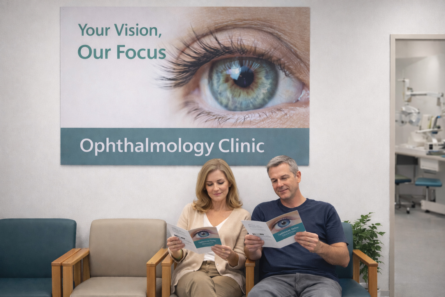
We offer dedicated materials for the waiting room: videos for monitors and TVs,
informational resources, and printed supports (desk folders), designed to prepare
patients and reinforce the value of the examination before the visit.
Continuity and long-term relationship
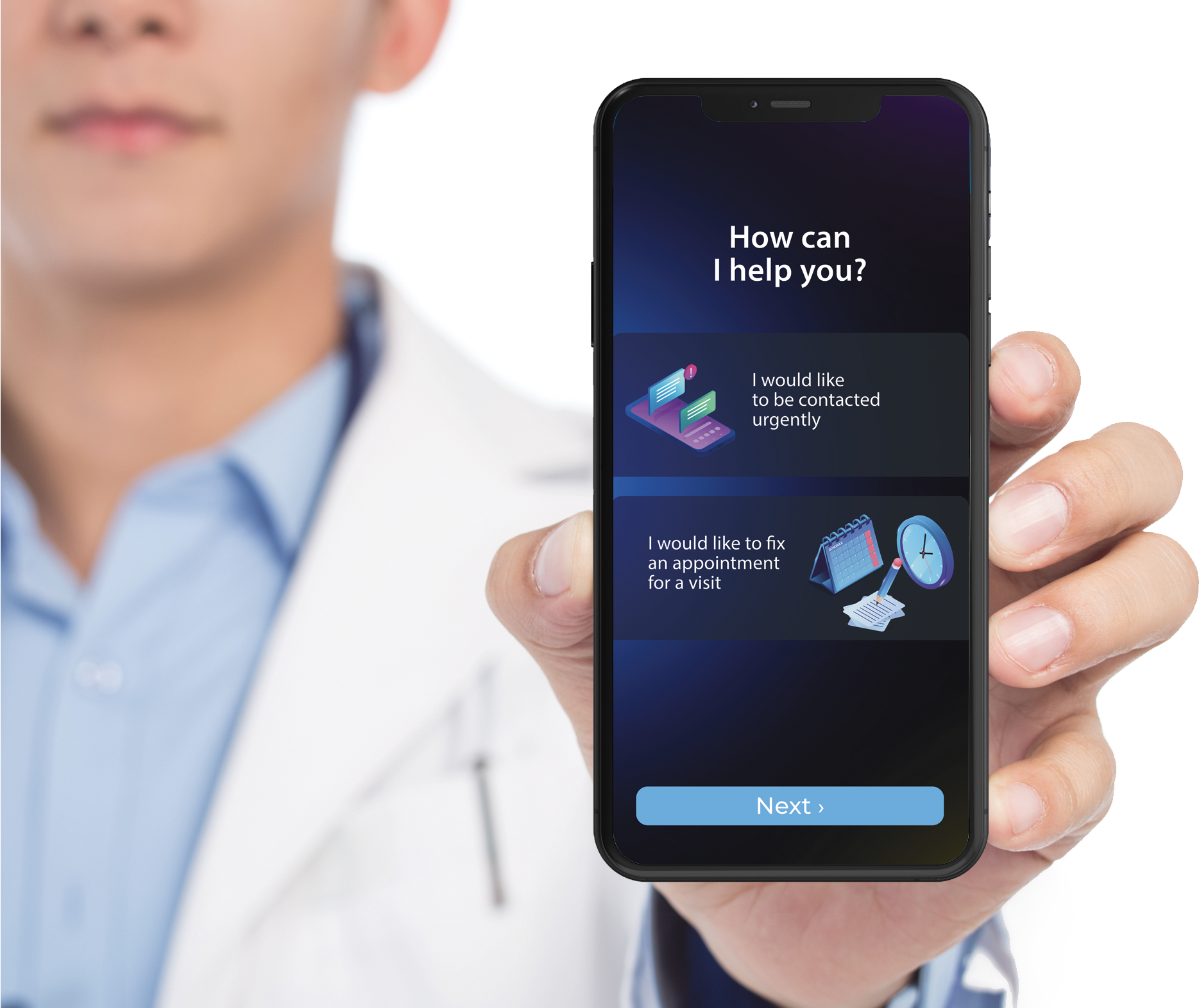
The Dry Eye Follow-Up software, provided free of charge, enables ongoing connection between patient
and professional, supporting follow-ups, reminders, and a continuous, structured care pathway.
The result is an integrated approach that reduces medical time, improves patient
understanding, and creates measurable clinical and economic value for the facility.
In-office services
Online awareness
Education about conditions
Patient preparation before the visit
No doctor time required
The patient arrives already motivated

In-clinic awareness
Dedicated and clear materials
Messaging aligned with the patient journey
Reinforcing the value of the diagnosis
The patient understands why the examination is important
Diagnosis
2 minutes – Fully automatic
Fast, objective, and standardized examination
No operator dependency
Easily delegated to staff
Relief
Relief aligned with the diagnosis
Clear pathway that can be easily explained to the patient
Clinical value + economic value
Retention and loyalty
Reminders via the “Dry Eye Follow-Up” app
and personalized recommendations for
a continuous care pathway.
SBM Dry Eye Clinical Report
From Diagnosis to Clinical Guidance
Many diagnostic devices generate images and measurements. SBM technology goes one step further.
At the end of the examination, the system automatically generates a Dry Eye Clinical Report,
summarizing the diagnostic findings and suggesting possible therapeutic approaches.
Transforming diagnostic data into clinical guidance.
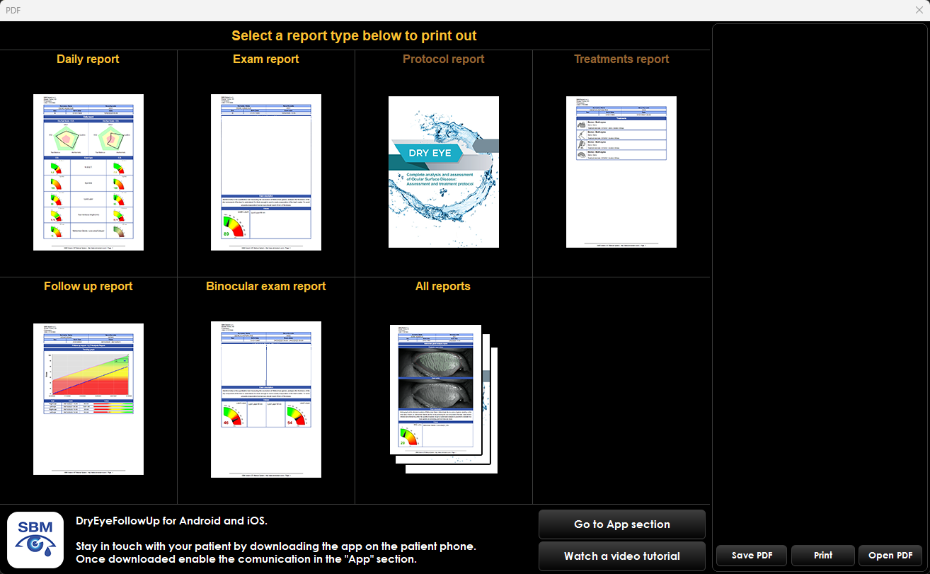
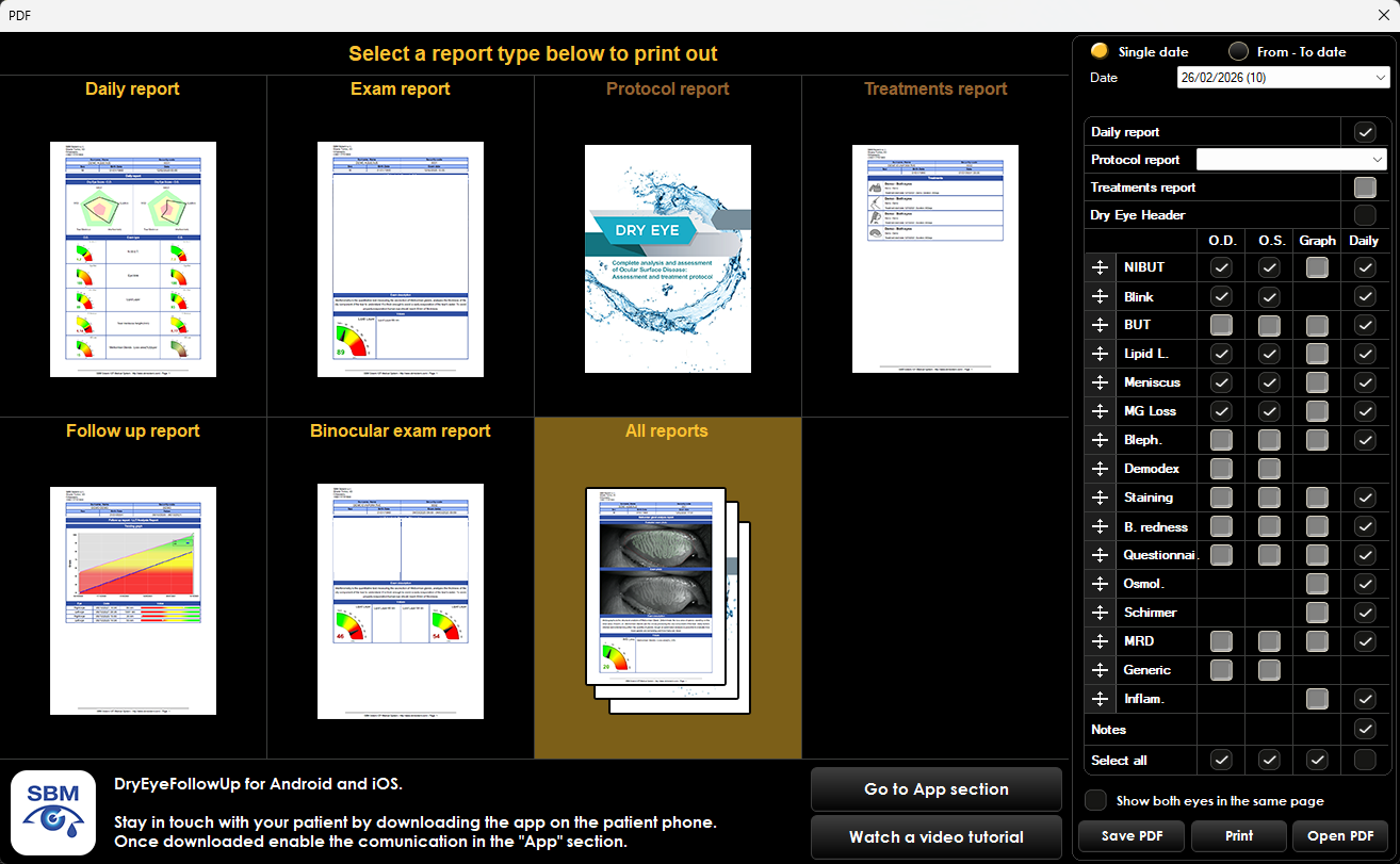
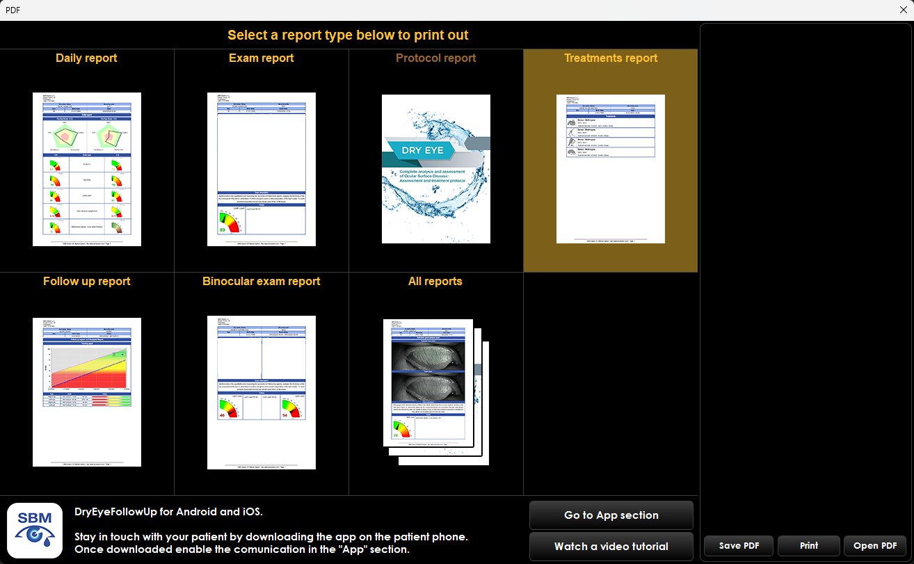
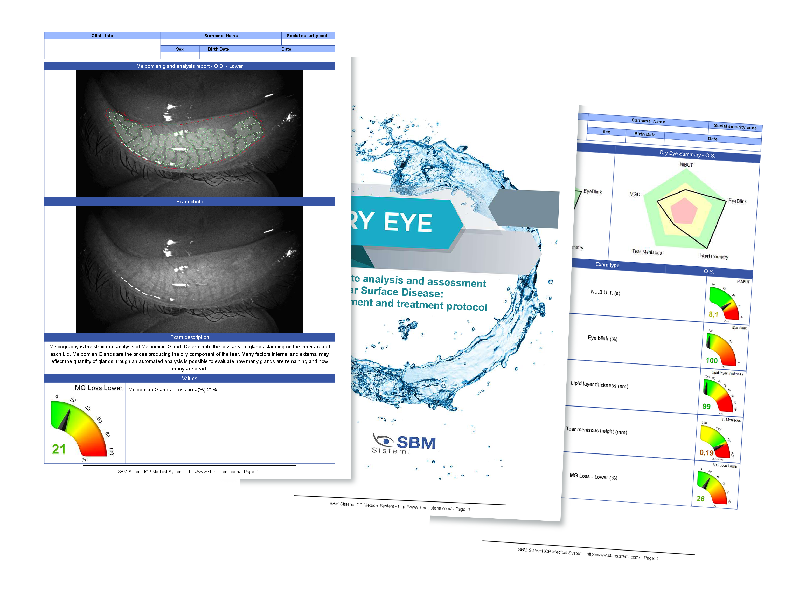
All reports
The ‘All Reports’ option allows printing a folder containing up to 16 pages. When the patient bears the costs personally, receiving a detailed, multi-page report represents added value and enhances satisfaction.
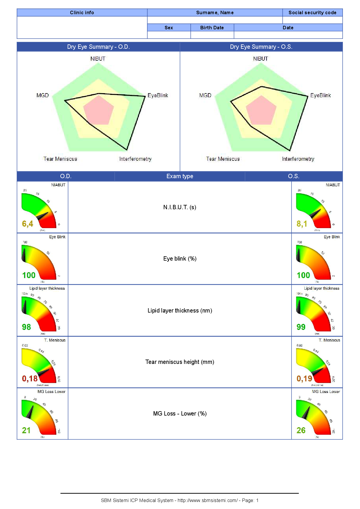
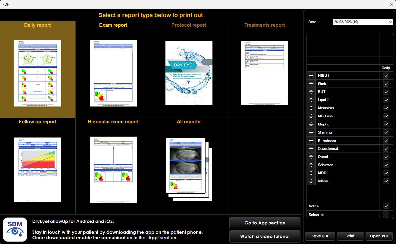
Daily report
The ‘Daily Report’ consolidates all results from a patient’s examinations performed on a single day. Various display options are available, with or without examination images.

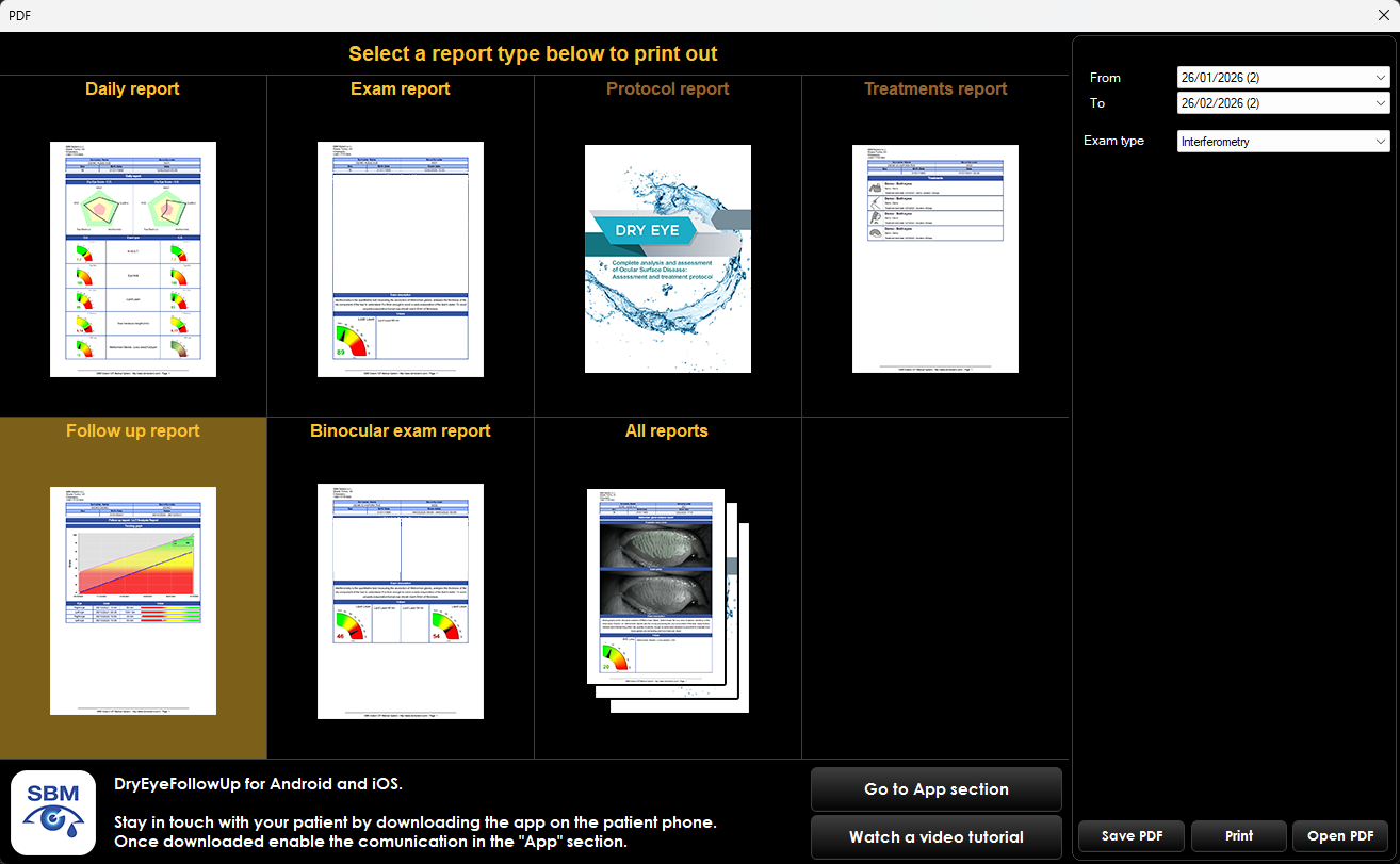
Follow-Up report
The ‘Follow-Up Report’ can show the patient the benefits
of therapy, highlighting improvements in symptoms
related to Dry Eye Disease.

Treatments Report
The ‘Treatments Report’ lists all treatments
prescribed to the patient.
SBM Sistemi not only measures the eye
But also helps guide therapeutic decisions
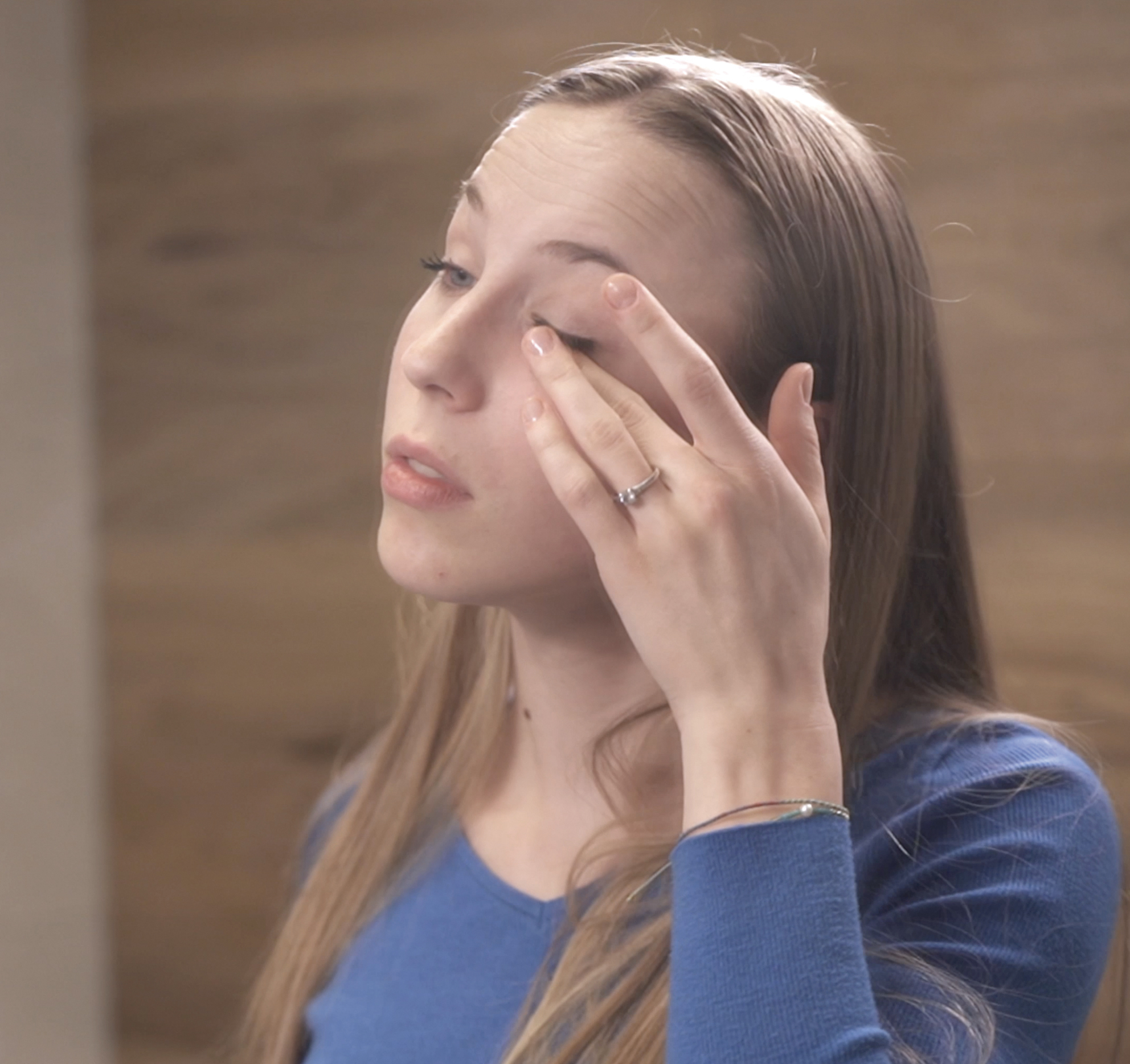
Patient symptoms
Capture and evaluate patient-reported discomfort, dryness, and visual disturbances.
Objective measurements
Provide precise, repeatable data on tear film and ocular surface condition.
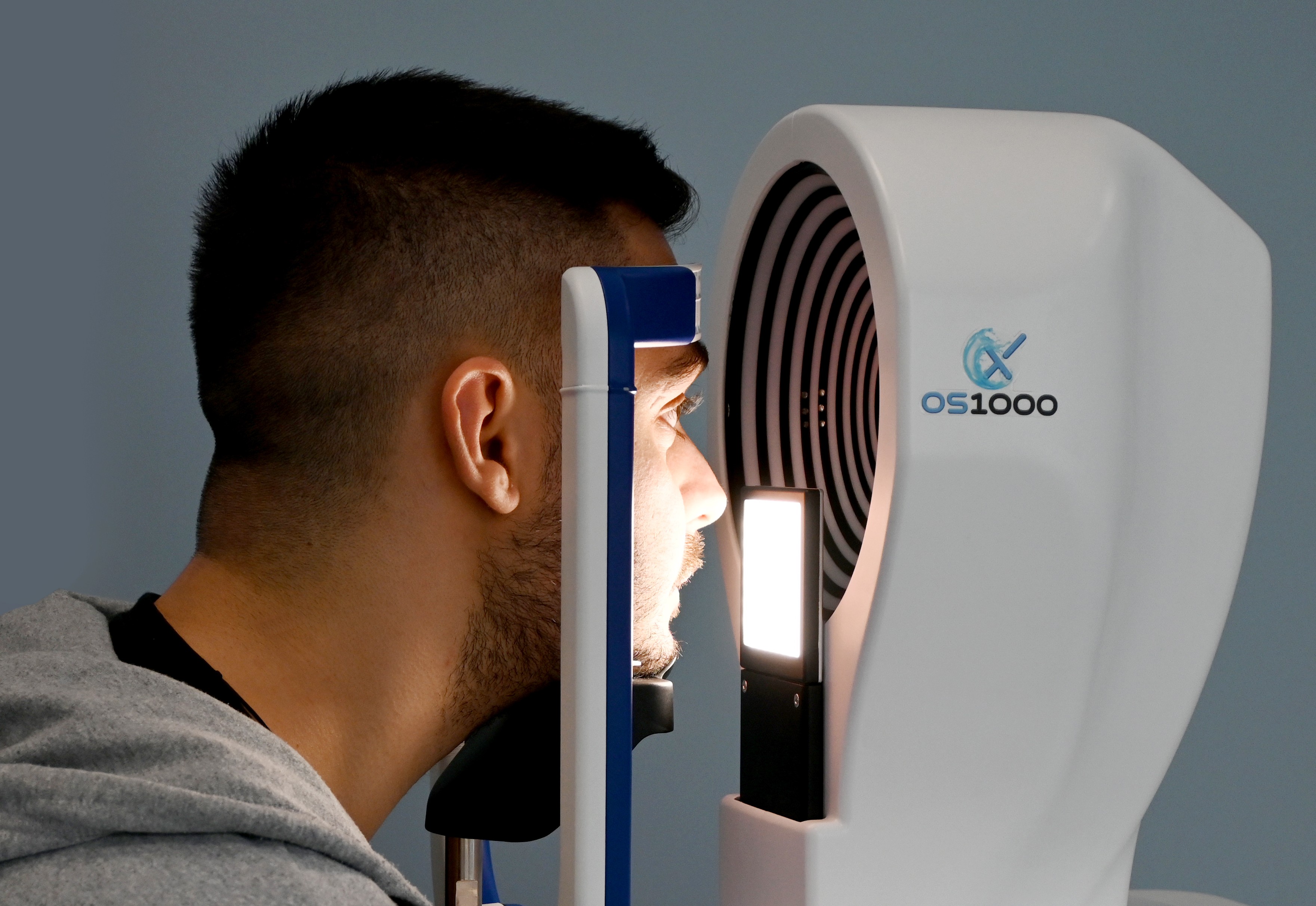
Dry Eye classification
Automatically categorize Dry Eye type and severity for accurate diagnosis.
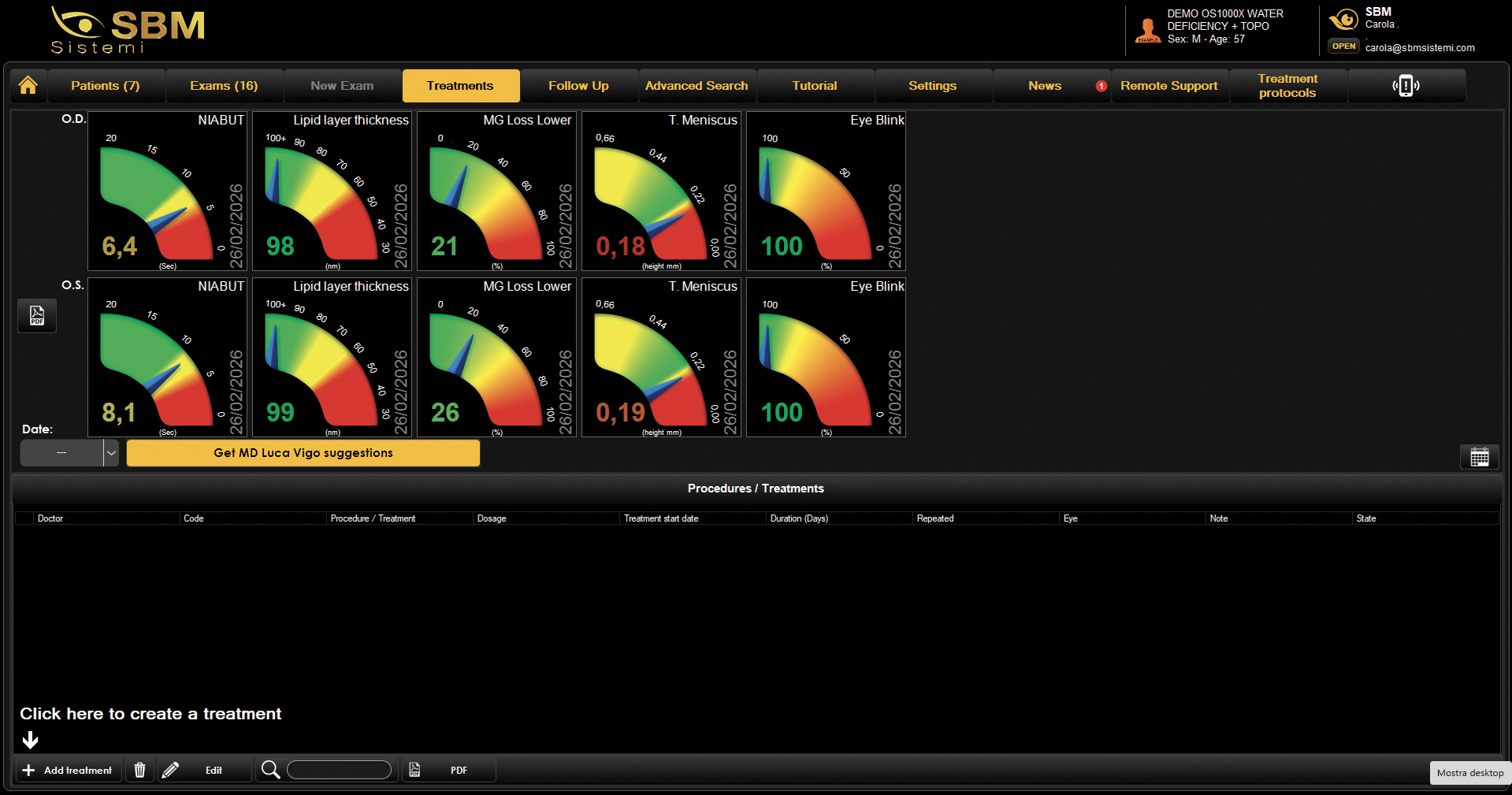
Treatment suggestions
Support clinical decision with tailored, evidence-based treatment options.
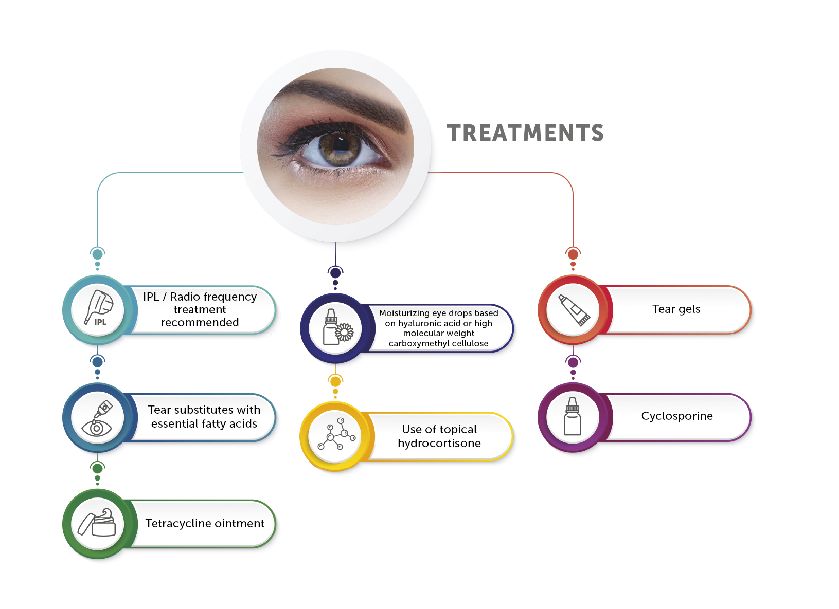
Clinical Advantages
Objective interpretation of results
Easier explanation to the patient
Support for treatment decisions
Standardized clinical workflow
Practice Advantages
Faster clinical decisions
Easier delegation to staff
Improved patient acceptance of treatment
Structured Dry Eye services
Most diagnostic systems provide data.
SBM technology provides clinical guidance.
Treatment protocol
The software offers multiple versatile solutions carefully designed to support medical
professionals and users in the complex task of assigning therapy.
Automatic
Treatment suggestion
The exclusive integrated algorithm, created in collaboration with Dr. Luca Vigo from
Studio Medico Carones, offers a personalized therapeutic approach based on the analysis results.
Customized
Automatic treatment assignment
Each user can customize their own protocol (making it possible to delegate the diagnosis to an assistant).
Each option provides complete, dedicated, and printable reports.
Manual
Treatment management
Using the Treatment Management tab, the software enables clinicians to compile a database of all
treatments available in the practice. Treatment brand and type can be entered into the system
and prescribed to patients with just two clicks.
Intended Use and Regulatory Information
Intended Users
The device is intended for use by qualified healthcare professionals trained in ophthalmology, optometry or related eye care disciplines.
Clinical Use
OS1000 provides measurements and images that may support clinical evaluation, contact lens fitting, corneal morphology assessment and ocular surface examination according to the professional judgement of the healthcare professional.
Classification
Class IIa CE marked according to Regulation (EU) 2017/745.
OS1000 X
Corneal Topographer


The device does not provide an autonomous diagnosis. All measurements, images and numerical values generated by the device must be interpreted by a qualified healthcare professional and shall not be used as the sole basis for diagnosis or treatment decisions.
The availability of specific measurements and software functions may depend on the selected device configuration

Attention: The information on this page is intended exclusively for qualified healthcare professionals.


