


Smart Studio
Smart Studio

Lens Effect 3D
Lens Effect 3D

Optic Studio
Optic Studio
Best non-invasive tests for Dry Eye diagnosis in 2026
March 12, 2026
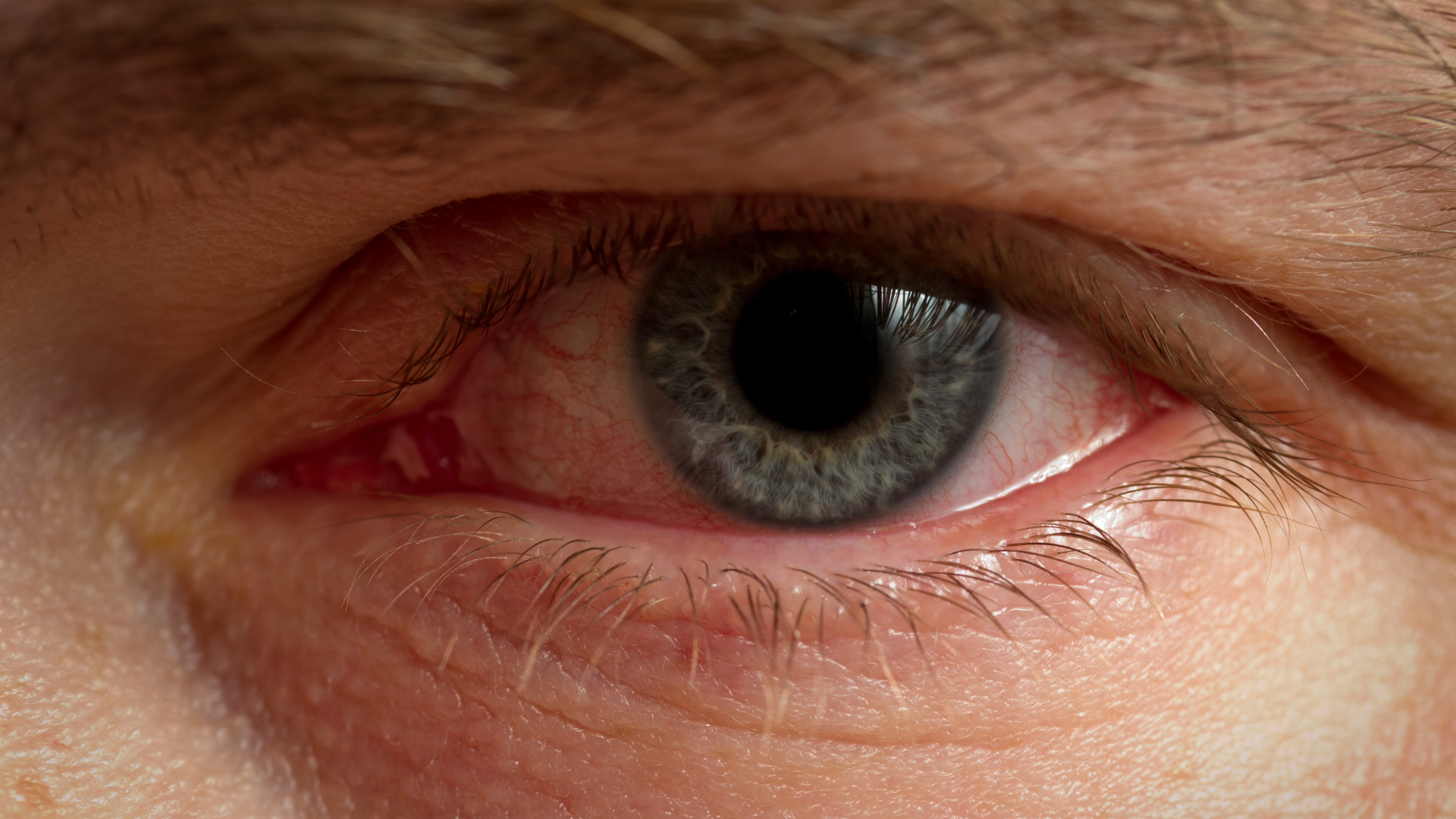
Dry eye disease (DED) continues to grow globally, driven by aging populations, digital device use, environmental
stressors, and refractive surgery.
In 2026, accurate diagnosis no longer relies on invasive dye-based techniques alone. Clinics are increasingly
adopting non-invasive dry eye diagnostic tests that provide objective, reproducible, and patient-friendly results.
For ophthalmologists, optometrists, and distributors evaluating advanced dry eye diagnostic devices, understanding
which technologies represent the current gold standard is essential.
Why non-invasive testing is the new standard
Traditional dry eye evaluation methods such as fluorescein TBUT and Schirmer test have well-known limitations:
- Reflex tearing affecting results
- Low repeatability
- Patient discomfort
- Subjective interpretation
Modern ocular surface analysis focuses on preserving physiological tear film conditions while extracting measurable
parameters.
Non-invasive tear film analysis enables:
- Greater reproducibility
- Standardized protocols
- Better treatment monitoring
- Higher patient acceptance
Integrated platforms such as IDRA X by SBM Sistemi illustrate this evolution in diagnostic workflow:
https://www.sbmsistemi.com/en/devices/idrax
Non-Invasive Tear Break-Up Time (NIBUT)
NIBUT measures tear film stability without fluorescein dye. Using projected ring patterns or Placido disc technology,
the system detects tear film disruption objectively.
Why it matters in 2026:
- Eliminates artificial destabilization caused by dye
- Improves repeatability
- Enables longitudinal patient tracking
- Enhances early evaporative dry eye detection
Clinics increasingly consider NIBUT a mandatory parameter in dry eye screening protocols.
Lipid Layer Thickness (LLT) measurement
Interferometry-based lipid layer thickness measurement analyzes the interference patterns created by the tear film’s
outer lipid layer.
This test is critical for diagnosing evaporative dry eye linked to Meibomian Gland Dysfunction (MGD).
Benefits:
- Non-contact evaluation
- Quantitative lipid grading
- Objective MGD classification
- Monitoring of treatment response
When combined with therapeutic solutions such as Activa:
https://www.sbmsistemi.com/en/devices/activa
clinicians can structure a complete MGD management pathway, from diagnosis to gland rehabilitation.
Infrared meibography
Infrared meibography visualizes meibomian gland morphology and dropout.
In 2026, structural gland analysis is considered essential because:
- MGD is one of the leading causes of evaporative dry eye
- Early gland loss can be asymptomatic
- Structural changes often precede severe symptoms
Quantitative meibography improves:
- Treatment timing
- Patient education
- Outcome monitoring
Integrated ocular surface analyzers that include meibography allow comprehensive dry eye evaluation in a single session.
Tear Meniscus Height measurement
Tear meniscus height (TMH) evaluates tear volume non-invasively using high-resolution imaging.
Clinical value:
- Detects aqueous-deficient dry eye
- Supports Sjögren screening
- Complements lipid layer analysis
- Avoids reflex tearing seen in Schirmer testing
This parameter is particularly useful when differentiating mixed dry eye forms.
Blink analysis and ocular surface dynamics
Modern dry eye diagnostic devices now include blink frequency and completeness analysis.
Why this matters:
- Incomplete blinking increases evaporation
- Digital device users show altered blink patterns
- Functional instability may occur even with normal tear production
By combining structural and functional parameters, clinicians obtain a more holistic view of ocular surface health.
Integrated ocular surface workstations: the 2026 approach
The real advancement in 2026 is not a single test, but integration.
Advanced platforms like IDRA X consolidate:
- NIBUT
- Lipid layer thickness
- Meibography
- Tear meniscus analysis
- Blink evaluation
into one unified system:
https://www.sbmsistemi.com/en/devices/idrax
This integrated approach improves:
- Workflow efficiency
- Diagnostic consistency
- Data comparability
- Patient communication
Clinical and economic impact for practices
Adopting non-invasive dry eye diagnostic tests allows clinics to:
- Differentiate from competitors
- Establish structured dry eye services
- Increase patient trust
- Improve conversion to advanced treatments
- Standardize pre- and post-therapy evaluations
When diagnostic imaging supports therapeutic solutions such as Activa, clinics can create complete dry eye treatment pathways.
What distributors should consider in 2026
For distributors evaluating ophthalmic device manufacturers, non-invasive diagnostic systems should offer:
- CE-certified production
- High-resolution imaging
- Modular scalability
- Software upgradability
- Long-term manufacturing reliability
SBM Sistemi combines decades of experience in ophthalmic device development with European manufacturing standards.
Learn more about the company’s background here:
https://www.sbmsistemi.com/en/company/history
Strong R&D capability and structured international support remain key factors for long-term distribution
partnerships.
In 2026, the best non-invasive tests for dry eye diagnosis are those that combine objectivity, repeatability, and
integration.
NIBUT, lipid layer interferometry, meibography, tear meniscus measurement, and blink analysis together define the
new gold standard.
Clinics and distributors seeking advanced dry eye diagnostic devices should prioritize integrated ocular surface
analyzers that deliver comprehensive, data-driven evaluation, improving both clinical outcomes and business
sustainability.
Read more
Objective vs subjective meibography: why quantification matters
Dry Eye in dogs and cats: beyond the Schirmer Test
The business case for Dry Eye clinics: revenue opportunities and patient retention
How corneal topography helps detect keratoconus in early stages
Automated Dry Eye diagnosis: how technology improves repeatability and workflow
Corneal Topography and Dry Eye: why both matter before cataract surgery
Tear film layers explained: lipid, aqueous and mucin
How to build a complete Dry Eye diagnostic workflow in your clinic
How to choose a Dry Eye diagnostic device for your practice
Meibomian gland dropout: early detection with advanced imaging
How objective Dry Eye diagnostics increase clinic revenue
Objective Optical Quality Metrics in Modern Ophthalmology
Regulatory Considerations When Importing CE Certified Ophthalmic Devices
Quantitative meibomian gland analysis: from images to objective data
OEM Opportunities in Ophthalmic Diagnostic Equipment
Transillumination meibography: clinical applications and advantages
What is a Tear Film Analyzer Device and how does it improve Dry Eye diagnosis?
Ocular surface assessment in high-volume clinics: efficiency without compromising accuracy
Beyond dry eye: other ocular surface disorders that impact visual quality
The role of ocular surface inflammation in post-surgical patient dissatisfaction
Ocular surface evaluation in contact lens wearers: what clinicians often miss
Environmental and lifestyle factors that alter tear film stability
Tear Film health in French Bulldogs: what the latest study using OSA-Vet® reveals
Pre-surgical dry eye screening: which parameters really influence outcomes
Optimizing the ocular surface before cataract surgery to improve surgical accuracy and patient outcomes
Dry eye and cataract surgery: managing expectations through objective preoperative evaluation
Evidence-based guidelines for cataract surgery: improving outcomes through standardized clinical practice
Meibomian gland dysfunction: why early detection changes long-term patient outcomes
Meibography and MGD: from gland imaging to clinical decision making
Non-invasive tear film assessment: clinical advantages over fluorescein-based methods
Tear film analysis explained: LLT, NIBUT and tear meniscus in clinical practice
Why traditional dry eye tests are not enough: limits of Schirmer and TBUT in modern practice
Dry eye diagnosis today: why ocular surface analysis is no longer optional

Attention: The information on this page is intended exclusively for qualified healthcare professionals.


