


Smart Studio
Smart Studio

Lens Effect 3D
Lens Effect 3D

Optic Studio
Optic Studio
Dry Eye in dogs and cats: beyond the Schirmer Test
July 22, 2026
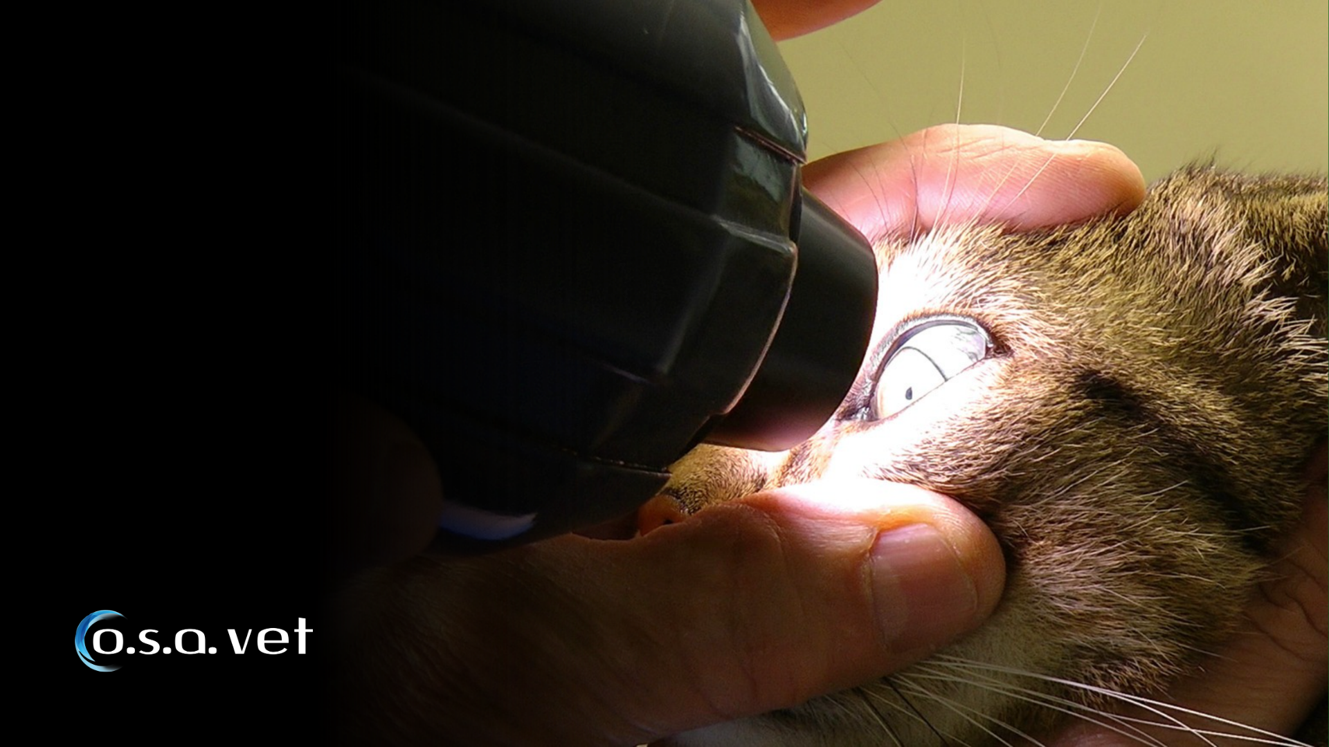
For decades, the Schirmer Tear Test (STT) has been considered the gold standard for diagnosing dry eye disease in
veterinary patients.
While tear production remains an essential component of ocular surface evaluation, our understanding of dry eye has
evolved significantly. Today, veterinary ophthalmologists recognize that many patients may present ocular surface
dysfunction even when Schirmer values fall within normal ranges.
Just as human ophthalmology has moved toward a comprehensive evaluation of the tear film and ocular surface,
veterinary eye care is beginning to adopt a broader diagnostic approach.
The challenge is no longer simply measuring tear quantity, but understanding tear quality, tear film stability,
blinking behavior, and meibomian gland health.
What Is Dry Eye disease in veterinary patients?
Dry eye disease, commonly referred to as Keratoconjunctivitis Sicca (KCS), is one of the most frequently encountered
ocular disorders in dogs.
The disease is characterized by insufficient tear film protection of the ocular surface, resulting in chronic
inflammation and discomfort.
Common clinical signs include:
- Ocular redness
- Mucoid discharge
- Excessive blinking
- Corneal pigmentation
- Corneal vascularization
- Recurrent corneal ulcers
- Reduced visual comfort
Although dogs are more commonly affected, cats can also develop tear film abnormalities associated with chronic ocular surface inflammation.
Why the Schirmer Tear Test is only part of the story
The Schirmer Tear Test remains an extremely valuable examination because it measures aqueous tear production.
However, tears are not composed exclusively of water.
The tear film is a complex structure consisting of:
- Lipid layer
- Aqueous layer
- Mucin layer
A patient may produce an adequate quantity of tears while still experiencing tear film instability and ocular surface
disease.
This concept is already well established in human dry eye diagnostics, as discussed in our article.
Increasingly, veterinary specialists are encountering cases where symptoms cannot be fully explained by Schirmer
values alone.
The growing importance of Meibomian Gland Dysfunction
Meibomian glands are responsible for producing the lipid layer of the tear film.
Their function is essential because the lipid layer:
- Reduces tear evaporation
- Stabilizes the tear film
- Protects the ocular surface
- Maintains visual quality
When meibomian gland dysfunction (MGD) occurs, excessive tear evaporation may develop even when tear production
appears normal.
In human ophthalmology, MGD is now recognized as one of the leading causes of dry eye disease.
Veterinary ophthalmology is beginning to observe similar patterns.
Meibography: seeing what the Schirmer Test cannot
Meibography allows clinicians to directly visualize the structure of the meibomian glands.
This non-invasive imaging technique can reveal:
- Gland shortening
- Gland distortion
- Partial gland loss
- Advanced gland dropout
These structural changes may help explain persistent symptoms in patients with apparently normal tear production.
For a deeper understanding of gland imaging and its clinical value, readers may also find useful our article.
The ability to visualize meibomian gland morphology represents one of the most significant advances in modern ocular
surface diagnostics.
Tear Film stability: an emerging diagnostic frontier
A healthy tear film must remain stable between blinks.
When the tear film breaks down too quickly, the ocular surface becomes exposed to environmental stress and
inflammation.
In human medicine, Non-Invasive Tear Break-Up Time (NIBUT) has become a widely adopted method for assessing tear
film stability.
The same principles may provide valuable insights in veterinary patients, particularly when traditional tests fail
to explain clinical symptoms.
Evaluating tear film behavior allows clinicians to move beyond static measurements and investigate how the ocular
surface functions in real-world conditions.
Blink analysis and ocular surface health
Blinking plays a critical role in maintaining tear film distribution.
Incomplete or altered blinking patterns may contribute to:
- Tear evaporation
- Ocular surface exposure /li>
- Tear film instability
Advanced ocular surface analysis systems can evaluate blink quality and frequency, providing additional information
about functional tear film performance.
Combining blink analysis with meibography and tear film assessment offers a more complete picture of ocular surface
health.
A new diagnostic approach in veterinary ophthalmology
Modern veterinary ocular surface evaluation should consider multiple factors rather than relying on a single test.
A comprehensive examination may include:
Tear production assessment
Schirmer Tear Test and conventional tear volume evaluation.
Meibomian gland imaging
Meibography to assess gland structure and detect gland loss.
Tear film stability analysis
Evaluation of tear film quality and evaporation-related dysfunction.
Ocular surface assessment
Documentation of redness, inflammation, corneal changes, and eyelid abnormalities.
Blink evaluation
Assessment of tear film distribution dynamics.
Together, these examinations provide a more complete understanding of the mechanisms underlying ocular surface
disease.
Technology supporting veterinary ocular surface diagnostics
As veterinary ophthalmology continues to evolve, specialized diagnostic platforms are becoming increasingly
important.
The OSA-Vet platform was specifically developed to support veterinary ocular surface evaluation through objective
and repeatable examinations.
By integrating multiple diagnostic tools into a single workflow, clinicians can investigate different aspects of
ocular surface disease rather than relying solely on tear quantity measurements.
This approach supports more informed clinical decisions and improves long-term patient monitoring.
From Dry Eye to ocular surface disease
One of the most significant changes occurring in both human and veterinary ophthalmology is the shift from the
concept of "dry eye" to the broader concept of "ocular surface disease."
This evolution recognizes that tear production, meibomian gland function, blinking behavior, eyelid health, and
ocular surface integrity are all interconnected.
Rather than asking only:
"How many tears is this patient producing?"
Clinicians are increasingly asking:
"How well is the entire ocular surface system functioning?"
The answer often requires much more than a Schirmer Tear Test.
The Schirmer Tear Test remains an essential component of veterinary dry eye diagnosis, but it should not be
considered the complete picture.
Modern understanding of ocular surface disease highlights the importance of evaluating tear quality, meibomian gland
function, tear film stability, and blinking behavior alongside tear production.
As technologies such as meibography and ocular surface analysis become more accessible to veterinary specialists,
the diagnosis of dry eye in dogs and cats is evolving toward a more comprehensive and objective approach.
The future of veterinary ocular surface care lies not only in measuring tears, but in understanding the entire tear
film ecosystem.
Read more
Objective vs subjective meibography: why quantification matters
The business case for Dry Eye clinics: revenue opportunities and patient retention
How corneal topography helps detect keratoconus in early stages
Automated Dry Eye diagnosis: how technology improves repeatability and workflow
Corneal Topography and Dry Eye: why both matter before cataract surgery
Tear film layers explained: lipid, aqueous and mucin
How to build a complete Dry Eye diagnostic workflow in your clinic
How to choose a Dry Eye diagnostic device for your practice
Meibomian gland dropout: early detection with advanced imaging
How objective Dry Eye diagnostics increase clinic revenue
Objective Optical Quality Metrics in Modern Ophthalmology
Regulatory Considerations When Importing CE Certified Ophthalmic Devices
Quantitative meibomian gland analysis: from images to objective data
OEM Opportunities in Ophthalmic Diagnostic Equipment
Best non-invasive tests for Dry Eye diagnosis in 2026
Transillumination meibography: clinical applications and advantages
What is a Tear Film Analyzer Device and how does it improve Dry Eye diagnosis?
Ocular surface assessment in high-volume clinics: efficiency without compromising accuracy
Beyond dry eye: other ocular surface disorders that impact visual quality
The role of ocular surface inflammation in post-surgical patient dissatisfaction
Ocular surface evaluation in contact lens wearers: what clinicians often miss
Environmental and lifestyle factors that alter tear film stability
Tear Film health in French Bulldogs: what the latest study using OSA-Vet® reveals
Pre-surgical dry eye screening: which parameters really influence outcomes
Optimizing the ocular surface before cataract surgery to improve surgical accuracy and patient outcomes
Dry eye and cataract surgery: managing expectations through objective preoperative evaluation
Evidence-based guidelines for cataract surgery: improving outcomes through standardized clinical practice
Meibomian gland dysfunction: why early detection changes long-term patient outcomes
Meibography and MGD: from gland imaging to clinical decision making
Non-invasive tear film assessment: clinical advantages over fluorescein-based methods
Tear film analysis explained: LLT, NIBUT and tear meniscus in clinical practice
Why traditional dry eye tests are not enough: limits of Schirmer and TBUT in modern practice
Dry eye diagnosis today: why ocular surface analysis is no longer optional

Attention: The information on this page is intended exclusively for qualified healthcare professionals.


