


Smart Studio
Smart Studio

Lens Effect 3D
Lens Effect 3D

Optic Studio
Optic Studio
Objective vs subjective meibography: why quantification matters
July 29, 2026
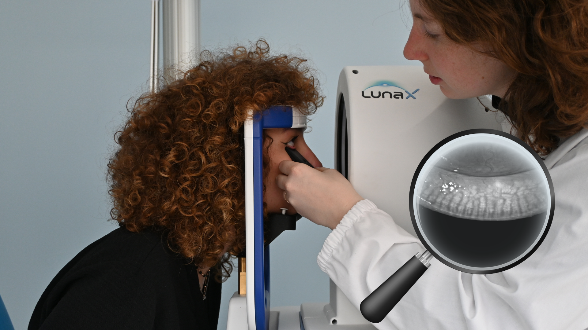
Meibography has become one of the most important diagnostic tools in modern dry eye management. As the understanding
of Meibomian Gland Dysfunction (MGD) continues to evolve, clinicians increasingly recognize that evaluating gland
structure is essential for understanding the underlying causes of evaporative dry eye disease.
Traditionally, meibography interpretation has relied largely on subjective assessment. Clinicians visually inspect
gland morphology and estimate the extent of gland loss based on personal experience and grading scales.
While this approach remains valuable, advances in imaging technology and software analysis are shifting the field
toward quantitative meibography, where gland morphology can be measured, documented, and monitored objectively over
time.
This evolution mirrors a broader trend in ophthalmology: moving from subjective observation toward standardized,
repeatable, and data-driven diagnostics.
Why Meibomian glands matter
The meibomian glands play a critical role in maintaining ocular surface health.
Located within the eyelids, these specialized glands produce the lipid layer of the tear film, which helps:
- Reduce tear evaporation
- Stabilize the tear film
- Protect the ocular surface
- Improve visual quality
When gland function becomes compromised, excessive evaporation may occur, leading to symptoms commonly associated
with dry eye disease.
Today, MGD is considered one of the leading causes of evaporative dry eye worldwide.
For a broader overview of modern dry eye diagnostics, readers may also find useful this article.
The traditional approach: subjective meibography
For many years, meibography interpretation has depended primarily on visual assessment.
The clinician evaluates infrared images and estimates:
- Gland dropout
- Gland shortening
- Gland distortion
- Gland tortuosity
- Overall gland morphology
Several grading systems have been proposed to classify meibomian gland loss, often assigning scores based on the
estimated percentage of gland dropout.
Although widely used, subjective grading presents several limitations:
Inter-observer variability
Different clinicians may assign different scores to the same image.
Intra-observer variability
Even the same clinician may grade the image differently at different time points.
Limited sensitivity
Small changes in gland morphology may not be easily detectable through visual estimation alone.
Difficult longitudinal monitoring
Tracking disease progression over months or years becomes challenging when assessments rely solely on subjective interpretation.
The growing need for quantitative meibography
As dry eye management becomes increasingly evidence-based, clinicians require more objective methods for evaluating
disease severity and treatment outcomes.
This has driven the development of quantitative meibography systems capable of measuring gland morphology rather
than simply estimating it.
Quantitative analysis can provide objective information regarding:
- Percentage of gland loss
- Gland area
- Gland density
- Morphological changes
- Structural asymmetry
By converting images into measurable data, clinicians gain access to more reproducible and comparable information.
How meibography software improves repeatability
Modern meibography software uses image processing algorithms to identify and analyze gland structures automatically.
Rather than relying entirely on visual interpretation, software-assisted analysis can:
- Standardize gland identification
- Reduce operator dependency
- Improve consistency
- Facilitate longitudinal follow-up
- Support research applications
This is particularly valuable when monitoring patients over extended periods.
Objective measurements allow clinicians to compare examinations performed months or years apart using the same
evaluation criteria.
The importance of quantification in MGD grading
One of the most challenging aspects of Meibomian Gland Dysfunction management is determining whether a patient is
improving, stable, or progressing.
Traditional MGD grading scales often rely on broad categories that may not capture subtle structural changes.
Quantitative meibography offers several advantages:
Better baseline documentation
Clinicians can establish objective reference values at the first visit.
Improved monitoring
Changes in gland morphology can be tracked more accurately over time.
Treatment evaluation
Objective measurements help assess the effectiveness of therapeutic interventions.
Enhanced patient communication
Patients often better understand their condition when presented with measurable data rather than subjective descriptions.
Beyond dropout: the evolution of gland analysis
Historically, most meibography systems focused primarily on gland dropout.
Today, clinicians are becoming increasingly interested in a wider range of structural parameters.
Advanced meibomian gland analysis may evaluate:
- Gland length
- Gland width
- Gland tortuosity
- Gland density
- Gland thickness
- Spatial distribution
These additional parameters may provide deeper insights into gland health and disease progression.
The future of MGD diagnosis will likely involve a combination of structural, functional, and quantitative
biomarkers.
The emergence of 3D meibography
One of the most exciting developments in ocular surface imaging is the introduction of three-dimensional gland
visualization.
Traditional meibography provides a two-dimensional representation of gland morphology.
Newer technologies aim to provide enhanced visualization of gland architecture, allowing clinicians to appreciate
structural details that may be difficult to identify using conventional imaging alone.
Three-dimensional visualization may improve:
- Morphological assessment
- Clinical documentation
- Patient education
- Research applications
As imaging technology continues to evolve, 3D meibography has the potential to become an increasingly valuable tool in ocular surface diagnostics.
Quantitative meibography and research applications
Research centers and key opinion leaders increasingly require objective metrics that can be compared across studies
and institutions.
Quantitative analysis provides several advantages for clinical research:
- Standardized measurements
- Improved reproducibility
- Reduced observer bias
- Better longitudinal analysis
- Enhanced data comparability
These characteristics make quantitative meibography particularly attractive for multicenter studies investigating dry eye disease and Meibomian Gland Dysfunction.
Integrating quantitative meibography into clinical practice
Meibography should not be considered an isolated examination.
Its greatest value emerges when integrated into a comprehensive ocular surface evaluation.
A complete diagnostic workflow may include:
- Meibography
- Interferometry
- Tear Meniscus Height
- Non-Invasive Break-Up Time (NIBUT)
- Blink Analysis
Together, these examinations provide a comprehensive understanding of tear film function and ocular surface health.
Clinicians interested in the broader role of meibomian gland imaging may also enjoy reading this previous article,
which explores how advanced diagnostics can support specialized dry eye services.
Technology driving objective gland analysis
The shift toward objective meibography is being accelerated by the availability of advanced diagnostic platforms.
Systems such as the IDRA X Dry Eye Analyzer and the OS1000 X Automated Corneal Topographer and Ocular Surface
Analyzer integrate meibography with multiple ocular surface examinations, supporting a more comprehensive diagnostic
workflow.
By combining automated acquisition, gland visualization, and advanced analysis tools, these platforms help
clinicians obtain objective information while maintaining workflow efficiency.
The future of Meibomian gland analysis
As dry eye diagnostics continue to evolve, the role of quantitative imaging will become increasingly important.
Future developments will likely focus on:
- Artificial intelligence-assisted analysis
- Automated gland segmentation
- Predictive progression models
- Advanced three-dimensional visualization
- Integrated multimodal ocular surface assessment
The objective is not simply to generate more data, but to provide clinically meaningful information that improves patient care.
Meibography has transformed the way clinicians evaluate Meibomian Gland Dysfunction, but subjective interpretation
alone may no longer be sufficient for modern dry eye management.
Quantitative meibography provides objective, repeatable, and measurable information that can improve diagnostic
consistency, facilitate longitudinal monitoring, and support both clinical practice and research.
As technologies such as automated gland analysis and 3D meibography continue to mature, objective evaluation of
gland morphology is likely to become an increasingly important component of ocular surface diagnostics.
For clinicians seeking greater precision in MGD grading and meibomian gland analysis, quantification is not simply a
technological enhancement—it represents the next step in the evolution of dry eye diagnosis.
Read more
Dry Eye in dogs and cats: beyond the Schirmer Test
The business case for Dry Eye clinics: revenue opportunities and patient retention
How corneal topography helps detect keratoconus in early stages
Automated Dry Eye diagnosis: how technology improves repeatability and workflow
Corneal Topography and Dry Eye: why both matter before cataract surgery
Tear film layers explained: lipid, aqueous and mucin
How to build a complete Dry Eye diagnostic workflow in your clinic
How to choose a Dry Eye diagnostic device for your practice
Meibomian gland dropout: early detection with advanced imaging
How objective Dry Eye diagnostics increase clinic revenue
Objective Optical Quality Metrics in Modern Ophthalmology
Regulatory Considerations When Importing CE Certified Ophthalmic Devices
Quantitative meibomian gland analysis: from images to objective data
OEM Opportunities in Ophthalmic Diagnostic Equipment
Best non-invasive tests for Dry Eye diagnosis in 2026
Transillumination meibography: clinical applications and advantages
What is a Tear Film Analyzer Device and how does it improve Dry Eye diagnosis?
Ocular surface assessment in high-volume clinics: efficiency without compromising accuracy
Beyond dry eye: other ocular surface disorders that impact visual quality
The role of ocular surface inflammation in post-surgical patient dissatisfaction
Ocular surface evaluation in contact lens wearers: what clinicians often miss
Environmental and lifestyle factors that alter tear film stability
Tear Film health in French Bulldogs: what the latest study using OSA-Vet® reveals
Pre-surgical dry eye screening: which parameters really influence outcomes
Optimizing the ocular surface before cataract surgery to improve surgical accuracy and patient outcomes
Dry eye and cataract surgery: managing expectations through objective preoperative evaluation
Evidence-based guidelines for cataract surgery: improving outcomes through standardized clinical practice
Meibomian gland dysfunction: why early detection changes long-term patient outcomes
Meibography and MGD: from gland imaging to clinical decision making
Non-invasive tear film assessment: clinical advantages over fluorescein-based methods
Tear film analysis explained: LLT, NIBUT and tear meniscus in clinical practice
Why traditional dry eye tests are not enough: limits of Schirmer and TBUT in modern practice
Dry eye diagnosis today: why ocular surface analysis is no longer optional

Attention: The information on this page is intended exclusively for qualified healthcare professionals.


