


Smart Studio
Smart Studio

Lens Effect 3D
Lens Effect 3D

Optic Studio
Optic Studio
Automated Dry Eye diagnosis: how technology improves repeatability and workflow
June 10, 2026
Dry eye disease is one of the most common conditions encountered in daily eye care practice. Despite its prevalence,
diagnosing and monitoring dry eye remains challenging due to the multifactorial nature of the disease and the
variability of many traditional diagnostic methods.
Historically, several dry eye assessments have relied heavily on operator experience, patient cooperation, and
subjective interpretation. This can lead to inconsistencies between examinations and make it difficult to compare
results over time.
Today, advances in imaging, software analysis, and automation are transforming the way clinicians evaluate the
ocular surface. Automated dry eye diagnosis improves repeatability, reduces operator dependency, and helps create a
more efficient workflow.
As discussed in our article about Dry Eye Diagnosis, obtaining objective and reproducible data is essential for
accurate diagnosis, treatment planning, and long-term patient monitoring.
Why repeatability matters in Dry Eye diagnosis
Dry eye disease is often chronic and progressive. For this reason, clinicians must be able to compare examinations
performed over weeks, months, or even years.
A diagnostic test is only valuable if it can provide consistent results under similar conditions.
Poor repeatability may lead to:
- Misclassification of disease severity
- Difficulty evaluating treatment effectiveness
- Variability between operators
- Reduced confidence in clinical decisions
- Challenges when monitoring patients over time
This is particularly important in practices managing large numbers of dry eye patients or offering advanced treatments for Meibomian Gland Dysfunction (MGD).
The limitations of traditional manual examinations
Many traditional dry eye assessments require significant operator involvement.
Examples include:
- Manual interpretation of tear film patterns
- Subjective grading of meibography images
- Visual estimation of tear meniscus height
- Fluorescein-based evaluations
- Manual documentation of findings
Even experienced clinicians may interpret results differently.
As patient expectations continue to increase, eye care professionals are seeking more objective and standardized
methods to evaluate ocular surface health.
For a deeper understanding of the key parameters involved in dry eye assessment, readers can explore our article on
Tear Film.
How automation is transforming Dry Eye evaluation
Modern ocular surface analyzers use advanced imaging systems and software algorithms to automatically capture,
process, and analyze diagnostic data.
Rather than relying exclusively on operator interpretation, automated systems provide objective measurements based
on standardized acquisition protocols.
Benefits include:
- Greater repeatability
- Reduced operator dependency
- Faster examinations
- Improved workflow efficiency
- Enhanced patient communication
- More reliable follow-up comparisons
Automation allows clinicians to focus more on patient care and less on manual data collection.
Automatic NIBUT: a more objective assessment of tear film stability
What Is NIBUT?
Non-Invasive Break-Up Time (NIBUT) is one of the most important indicators of tear film stability.
It measures the time required for the tear film to begin breaking after a blink without using fluorescein dye.
The challenge of manual evaluation
Traditional assessment methods often require the clinician to visually identify the first signs of tear film
disruption.
This introduces subjectivity and may affect repeatability.
The benefits of automatic NIBUT
Automated NIBUT analysis allows the system to:
- Detect tear film break-up automatically
- Measure break-up times objectively
- Reduce operator influence
- Improve consistency between visits
Objective tear film analysis is becoming increasingly important in modern ocular surface evaluation.
Meibography automation: improving Meibomian gland assessment
Meibomian gland dysfunction is recognized as one of the leading causes of evaporative dry eye.
Meibography allows clinicians to visualize gland morphology and identify gland loss, shortening, distortion, and
atrophy.
Traditional Challenges
Manual gland assessment often depends on:
- Subjective grading scales
- Operator experience
- Visual estimation of gland loss
This can create variability between examinations and between different clinicians.
The advantages of automated meibography
Automated meibography systems can:
- Automatically detect gland structures
- Quantify gland loss
- Generate objective grading scores
- Improve examination repeatability
- Facilitate longitudinal patient monitoring
For additional insights into gland morphology and dysfunction, readers can refer to our article The Importance of Meibomian Gland Evaluation in Dry Eye Disease.
Automated interferometry and lipid layer analysis
The lipid layer plays a crucial role in preventing tear evaporation and maintaining ocular surface stability.
Modern dry eye analyzers can automatically evaluate interferometric patterns and lipid layer characteristics.
Automation helps clinicians:
- Standardize image acquisition
- Reduce interpretation variability
- Monitor treatment effectiveness
- Better understand Meibomian gland functionality
Combining lipid layer assessment with meibography and NIBUT creates a more comprehensive picture of ocular surface health.
Automated blink analysis: an often overlooked parameter
Blink quality and blink completeness directly influence tear film distribution.
Incomplete blinking may contribute to:
- Tear film instability
- Increased evaporation
- Meibomian gland dysfunction
- Contact lens discomfort
Automated blink analysis enables clinicians to evaluate:
- Blink frequency
- Blink completeness
- Blink dynamics
These objective measurements provide valuable information that may otherwise be difficult to assess consistently.
How automation improves clinical workflow
Beyond diagnostic accuracy, automation significantly improves operational efficiency.
Automated acquisition protocols help reduce examination time and simplify training requirements.
Benefits include:
- Faster patient throughput
- Reduced staff workload
- Standardized examination protocols
- Improved consistency across multiple operators
- Easier implementation in multi-location practices
This is particularly important as dry eye clinics continue to grow and patient demand increases.
A more effective patient communication tool
One of the most valuable advantages of automated ocular surface analysis is the ability to generate objective visual
reports.
Patients can clearly see:
- Meibomian gland images
- Tear film measurements
- Lipid layer evaluations
- Blink analysis results
Visual evidence often improves patient understanding and treatment compliance.
Learn more about patient education and monitoring.
How IDRA X and OS1000 X support automated Dry Eye diagnosis
Modern eye care professionals increasingly seek diagnostic solutions that provide objective, repeatable, and
efficient examinations.
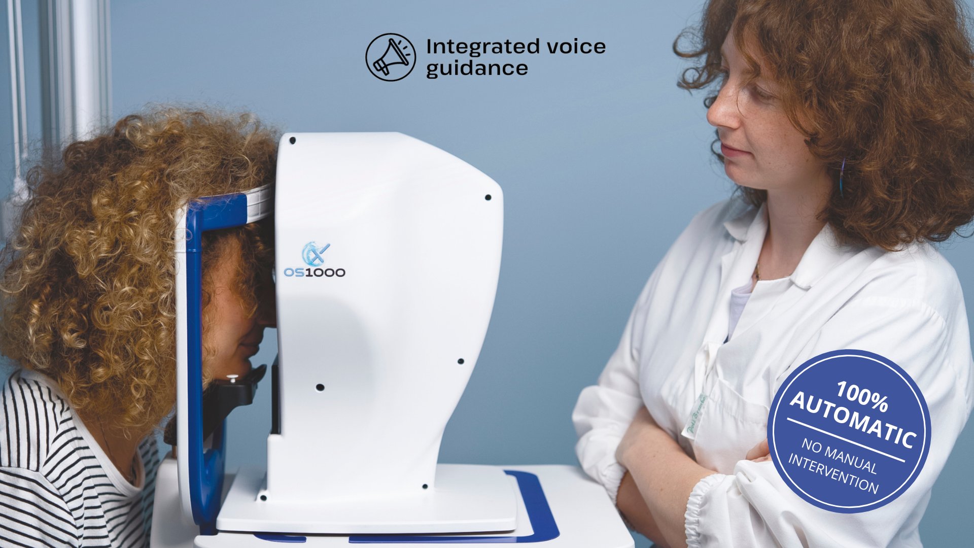
Both the IDRA X and OS1000 X integrate advanced dry eye evaluation tools designed to support standardized ocular
surface assessment.
These platforms include:
- Automatic NIBUT analysis
- Automated meibography
- Interferometry
- Tear meniscus assessment
- Blink analysis
- Ocular redness evaluation
In addition, the OS1000 X combines comprehensive dry eye evaluation with advanced corneal topography, allowing clinicians to assess both ocular surface health and corneal integrity within a single workflow.
Conclusion
The future of dry eye diagnosis is increasingly driven by automation.
Automated technologies help improve repeatability, reduce operator dependency, standardize examinations, and enhance
clinical efficiency.
By combining objective measurements such as automatic NIBUT, automated meibography, interferometry, and blink
analysis, clinicians can achieve a more comprehensive understanding of ocular surface health while improving patient
management and long-term follow-up.
Integrated diagnostic platforms such as IDRA X and OS1000 X are helping practices move toward a more standardized,
efficient, and data-driven approach to dry eye care.
Read more
Objective vs subjective meibography: why quantification matters
Dry Eye in dogs and cats: beyond the Schirmer Test
The business case for Dry Eye clinics: revenue opportunities and patient retention
How corneal topography helps detect keratoconus in early stages
Corneal Topography and Dry Eye: why both matter before cataract surgery
Tear film layers explained: lipid, aqueous and mucin
How to build a complete Dry Eye diagnostic workflow in your clinic
How to choose a Dry Eye diagnostic device for your practice
Meibomian gland dropout: early detection with advanced imaging
How objective Dry Eye diagnostics increase clinic revenue
Objective Optical Quality Metrics in Modern Ophthalmology
Regulatory Considerations When Importing CE Certified Ophthalmic Devices
Quantitative meibomian gland analysis: from images to objective data
OEM Opportunities in Ophthalmic Diagnostic Equipment
Best non-invasive tests for Dry Eye diagnosis in 2026
Transillumination meibography: clinical applications and advantages
What is a Tear Film Analyzer Device and how does it improve Dry Eye diagnosis?
Ocular surface assessment in high-volume clinics: efficiency without compromising accuracy
Beyond dry eye: other ocular surface disorders that impact visual quality
The role of ocular surface inflammation in post-surgical patient dissatisfaction
Ocular surface evaluation in contact lens wearers: what clinicians often miss
Environmental and lifestyle factors that alter tear film stability
Tear Film health in French Bulldogs: what the latest study using OSA-Vet® reveals
Pre-surgical dry eye screening: which parameters really influence outcomes
Optimizing the ocular surface before cataract surgery to improve surgical accuracy and patient outcomes
Dry eye and cataract surgery: managing expectations through objective preoperative evaluation
Evidence-based guidelines for cataract surgery: improving outcomes through standardized clinical practice
Meibomian gland dysfunction: why early detection changes long-term patient outcomes
Meibography and MGD: from gland imaging to clinical decision making
Non-invasive tear film assessment: clinical advantages over fluorescein-based methods
Tear film analysis explained: LLT, NIBUT and tear meniscus in clinical practice
Why traditional dry eye tests are not enough: limits of Schirmer and TBUT in modern practice
Dry eye diagnosis today: why ocular surface analysis is no longer optional

Attention: The information on this page is intended exclusively for qualified healthcare professionals.


