


Smart Studio
Smart Studio

Lens Effect 3D
Lens Effect 3D

Optic Studio
Optic Studio
Tear film layers explained: lipid, aqueous and mucin
May 20, 2026
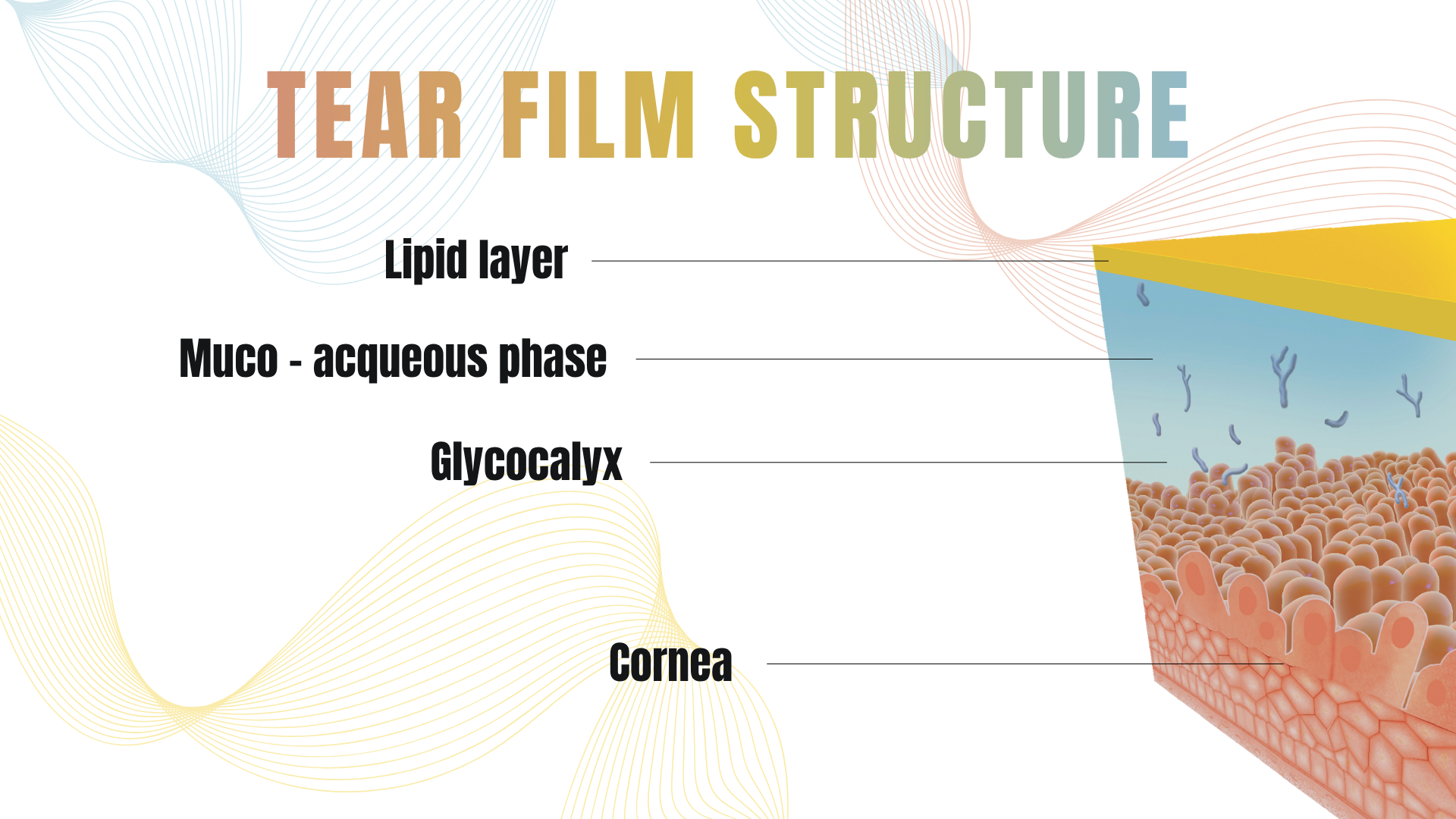
The tear film plays a fundamental role in maintaining ocular surface health, visual quality, and patient comfort.
Despite its microscopic thickness, it is a highly complex and dynamic structure composed of three distinct layers:
lipid, aqueous, and mucin.
Understanding how these tear film layers interact is essential for diagnosing and managing dry eye disease. Each
layer contributes to tear film stability, and dysfunction in any of them can lead to symptoms such as irritation,
fluctuating vision, and ocular discomfort.
In clinical practice, analyzing tear film layers has become increasingly important, especially with the availability
of advanced diagnostic technologies that allow non-invasive and objective assessment.
What is the tear film?
The tear film is a thin, multi-layered structure that covers the ocular surface. Its primary functions include:
- Maintaining optical clarity
- Protecting the cornea from environmental stress
- Providing nutrients and oxygen
- Supporting a smooth refractive surface
A stable tear film is essential for consistent vision. Even small disruptions in its structure can result in visual fluctuations and discomfort.
Overview of tear film layers
Traditionally, the tear film is described as consisting of three layers:
- Lipid layer (outer)
- Aqueous layer (middle)
- Mucin layer (inner)
Although modern research suggests a more integrated structure, this layered model remains clinically useful for understanding tear film dysfunction and guiding diagnosis.
The lipid layer: preventing evaporation
The outermost layer of the tear film is the lipid layer, produced by the meibomian glands. Its main function is to
reduce tear evaporation and stabilize the tear film.
A healthy lipid layer helps maintain tear film integrity between blinks. When this layer is compromised, evaporation
increases, leading to tear film instability and symptoms of evaporative dry eye.
Lipid layer alterations are commonly associated with meibomian gland dysfunction, one of the leading causes of dry
eye disease. Structural changes in the glands, including early signs of meibomian gland dropout, can significantly affect lipid production
and tear film quality.
Clinically, evaluating lipid layer thickness provides valuable insight into tear film performance and is a key
parameter in modern dry eye diagnostics.
The aqueous layer: hydration and nourishment
The aqueous layer is the thickest component of the tear film and is produced mainly by the lacrimal glands. It is responsible for:
- Hydrating the ocular surface
- Delivering nutrients and oxygen
- Removing debris and inflammatory mediators
A deficiency in the aqueous layer leads to aqueous-deficient dry eye, which is often associated with reduced tear
volume.
Assessing tear quantity through parameters such as tear meniscus height helps identify patients with reduced aqueous
production. When combined with other measurements, this contributes to a more complete ocular surface evaluation.
The mucin layer: ensuring tear film adhesion
The innermost layer of the tear film is the mucin layer, produced by goblet cells in the conjunctiva. Its primary
role is to ensure that the tear film adheres properly to the ocular surface.
Without mucin, the aqueous layer would not spread evenly across the cornea, leading to tear film instability and
localized dry spots.
Mucin deficiency is often associated with ocular surface damage, inflammation, or chronic dry eye conditions.
Although it is more difficult to measure directly compared to other layers, its role is critical in maintaining tear
film uniformity.
How tear film layers work together
Although described separately, the three tear film layers function as an integrated system.
- The mucin layer anchors the tear film to the ocular surface
- The aqueous layer provides hydration and nutrients
- The lipid layer protects against evaporation
Disruption in any one layer can destabilize the entire tear film. For example:
- Lipid deficiency increases evaporation
- Aqueous deficiency reduces tear volume
- Mucin deficiency prevents proper tear distribution
This interdependence explains why dry eye disease is often multifactorial and requires a comprehensive diagnostic approach.
Tear film instability and dry eye disease
Tear film instability is one of the key features of dry eye disease. It occurs when the balance between tear
production, distribution, and evaporation is disrupted.
One of the most widely used parameters to assess tear film stability is non-invasive break-up time (NIBUT), which
measures how long the tear film remains stable after a blink.
Modern diagnostic systems allow clinicians to evaluate tear film behavior dynamically and non-invasively, improving
both accuracy and repeatability compared to traditional methods.
Why understanding tear film layers matters in clinical practice
A detailed understanding of tear film layers helps clinicians:
- Identify the underlying cause of dry eye
- Differentiate between evaporative and aqueous-deficient forms
- Select appropriate treatment strategies
- Monitor disease progression over time
Rather than treating dry eye as a single condition, clinicians can tailor therapy based on which layer is primarily
affected.
This approach is increasingly supported by objective diagnostic workflows that integrate multiple parameters into a
comprehensive ocular surface assessment.
From theory to diagnosis: evaluating tear film layers
In modern ophthalmology, tear film evaluation goes beyond basic observation. Advanced diagnostic tools allow clinicians to assess:
- Lipid layer thickness
- Tear film stability (NIBUT)
- Tear meniscus height
- Meibomian gland structure
By combining these measurements, it becomes possible to build a complete picture of tear film function and identify
the dominant mechanism of dry eye.
This structured approach is at the core of an effective dry eye diagnostic workflow.
The tear film is a complex and dynamic system where lipid, aqueous, and mucin layers work together to maintain ocular
surface health and visual quality.
Understanding these layers is essential for accurate diagnosis and effective management of dry eye disease. As
diagnostic technologies continue to evolve, clinicians can rely on more objective and detailed assessments of tear
film function.
By integrating tear film analysis into routine practice, it becomes possible to move from symptom-based evaluation
to a more precise, data-driven approach to ocular surface care.
Read more
Objective vs subjective meibography: why quantification matters
Dry Eye in dogs and cats: beyond the Schirmer Test
The business case for Dry Eye clinics: revenue opportunities and patient retention
How corneal topography helps detect keratoconus in early stages
Automated Dry Eye diagnosis: how technology improves repeatability and workflow
Corneal Topography and Dry Eye: why both matter before cataract surgery
How to build a complete Dry Eye diagnostic workflow in your clinic
How to choose a Dry Eye diagnostic device for your practice
Meibomian gland dropout: early detection with advanced imaging
How objective Dry Eye diagnostics increase clinic revenue
Objective Optical Quality Metrics in Modern Ophthalmology
Regulatory Considerations When Importing CE Certified Ophthalmic Devices
Quantitative meibomian gland analysis: from images to objective data
OEM Opportunities in Ophthalmic Diagnostic Equipment
Best non-invasive tests for Dry Eye diagnosis in 2026
Transillumination meibography: clinical applications and advantages
What is a Tear Film Analyzer Device and how does it improve Dry Eye diagnosis?
Ocular surface assessment in high-volume clinics: efficiency without compromising accuracy
Beyond dry eye: other ocular surface disorders that impact visual quality
The role of ocular surface inflammation in post-surgical patient dissatisfaction
Ocular surface evaluation in contact lens wearers: what clinicians often miss
Environmental and lifestyle factors that alter tear film stability
Tear Film health in French Bulldogs: what the latest study using OSA-Vet® reveals
Pre-surgical dry eye screening: which parameters really influence outcomes
Optimizing the ocular surface before cataract surgery to improve surgical accuracy and patient outcomes
Dry eye and cataract surgery: managing expectations through objective preoperative evaluation
Evidence-based guidelines for cataract surgery: improving outcomes through standardized clinical practice
Meibomian gland dysfunction: why early detection changes long-term patient outcomes
Meibography and MGD: from gland imaging to clinical decision making
Non-invasive tear film assessment: clinical advantages over fluorescein-based methods
Tear film analysis explained: LLT, NIBUT and tear meniscus in clinical practice
Why traditional dry eye tests are not enough: limits of Schirmer and TBUT in modern practice
Dry eye diagnosis today: why ocular surface analysis is no longer optional

Attention: The information on this page is intended exclusively for qualified healthcare professionals.


