


Smart Studio
Smart Studio

Lens Effect 3D
Lens Effect 3D

Optic Studio
Optic Studio
Meibomian gland dropout: early detection with advanced imaging
April 29, 2026
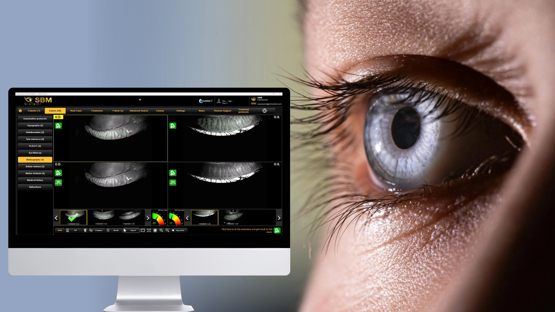
Meibomian gland dysfunction (MGD) is one of the leading causes of dry eye disease, yet it is often underdiagnosed in
its early stages. One of the most critical indicators of disease progression is meibomian gland dropout, a
structural alteration that can significantly impact tear film stability and ocular surface health.
Today, meibomian gland dropout imaging plays a key role in identifying these changes early, allowing clinicians to
intervene before irreversible damage occurs.
What Is Meibomian gland dropout?
Meibomian gland dropout refers to the partial or complete loss of meibomian glands within the eyelids. These glands
are responsible for producing the lipid layer of the tear film, which prevents evaporation.
When gland structure deteriorates, patients may experience:
- Tear film instability
- Increased evaporation
- Ocular discomfort
- Fluctuating vision
Because these structural changes can begin before symptoms become severe, relying only on patient complaints often leads to delayed diagnosis.
Why early detection is critical in MGD
Early-stage MGD is often reversible or manageable, while advanced gland dropout is not. This makes early detection
essential for both clinical outcomes and long-term patient management.
Modern approaches emphasize the importance of combining symptom evaluation with objective testing. As discussed in
SBM’s overview of non-invasive dry eye tests, reproducible and patient-friendly diagnostics are fundamental for
identifying dysfunction before it progresses.
By detecting gland alterations early, clinicians can:
- Initiate targeted treatment sooner
- Prevent disease progression
- Improve long-term tear film stability
The role of Meibomian gland dropout Imaging
Traditional slit lamp examination provides limited insight into gland morphology. In contrast, Meibomian gland
dropout imaging allows direct visualization of gland structure through meibography.
This technology makes it possible to:
- Assess gland loss and distortion
- Classify severity of MGD
- Monitor progression over time
Devices that integrate tear film analysis and meibography provide a more complete understanding of ocular surface health. For example, platforms like Idra X combine multiple diagnostic tools into a single workflow, allowing clinicians to correlate gland structure with tear film function.
From morphology to clinical decision-making
One of the biggest advantages of advanced imaging is its ability to connect structure with treatment decisions.
When clinicians can clearly visualize gland dropout, they can:
- Differentiate between evaporative and aqueous-deficient dry eye
- Personalize treatment strategies
- Set realistic expectations with patients
This aligns with the broader concept of ocular surface analysis, where multiple parameters are evaluated together
rather than in isolation.
For a deeper understanding of how imaging fits into the diagnostic process, see SBM’s explanation of the tear film
analyzer device and its role in comprehensive dry eye assessment.
Monitoring disease progression over time
Meibomian gland dropout is not only a diagnostic marker but also a tool for long-term monitoring.
By capturing baseline images and comparing them over time, clinicians can:
- Evaluate treatment effectiveness
- Track disease progression
- Adjust therapy protocols
This is particularly valuable in structured clinical workflows, such as those described in the article on ocular surface assessment in high-volume clinics, where efficiency and repeatability are essential.
Improving patient communication and treatment acceptance
Visual evidence plays a crucial role in patient education. Showing patients their gland structure helps them
understand the severity of their condition and the importance of treatment.
Instead of abstract explanations, clinicians can present clear images that demonstrate gland loss or alteration.
This significantly improves treatment acceptance and compliance.
When imaging is combined with treatment solutions such as Activa, patients can better understand the connection
between diagnosis and therapy, making the clinical pathway more coherent and convincing.
Integrating imaging into a Dry Eye workflow
To maximize its value, meibomian gland dropout imaging should be integrated into a broader diagnostic workflow.
A complete dry eye pathway includes:
- Symptom assessment
- Tear film analysis
- Meibography
- Treatment planning
- Follow-up monitoring
This integrated approach transforms dry eye from a reactive condition into a proactively managed disease.
Clinics that adopt structured workflows and advanced imaging technologies are better positioned to deliver
consistent results and improve patient satisfaction.
Clinical and business benefits of advanced imaging
Beyond clinical accuracy, imaging technologies also provide tangible benefits for clinic performance.
They help:
- Reduce diagnostic uncertainty
- Improve workflow efficiency
- Increase treatment acceptance
- Support recurring visits
By making dry eye more measurable and understandable, imaging contributes to both better care and stronger practice growth.
Meibomian gland dropout is a critical factor in dry eye disease progression, and early detection is essential for
effective management.
With the support of Meibomian gland dropout imaging, clinicians can move beyond subjective evaluation and adopt a
more precise, data-driven approach to ocular surface disease.
In today’s ophthalmic landscape, advanced imaging is not just a diagnostic upgrade, it is a fundamental component of
modern dry eye care.
Read more
Objective vs subjective meibography: why quantification matters
Dry Eye in dogs and cats: beyond the Schirmer Test
The business case for Dry Eye clinics: revenue opportunities and patient retention
How corneal topography helps detect keratoconus in early stages
Automated Dry Eye diagnosis: how technology improves repeatability and workflow
Corneal Topography and Dry Eye: why both matter before cataract surgery
Tear film layers explained: lipid, aqueous and mucin
How to build a complete Dry Eye diagnostic workflow in your clinic
How to choose a Dry Eye diagnostic device for your practice
How objective Dry Eye diagnostics increase clinic revenue
Objective Optical Quality Metrics in Modern Ophthalmology
Regulatory Considerations When Importing CE Certified Ophthalmic Devices
Quantitative meibomian gland analysis: from images to objective data
OEM Opportunities in Ophthalmic Diagnostic Equipment
Best non-invasive tests for Dry Eye diagnosis in 2026
Transillumination meibography: clinical applications and advantages
What is a Tear Film Analyzer Device and how does it improve Dry Eye diagnosis?
Ocular surface assessment in high-volume clinics: efficiency without compromising accuracy
Beyond dry eye: other ocular surface disorders that impact visual quality
The role of ocular surface inflammation in post-surgical patient dissatisfaction
Ocular surface evaluation in contact lens wearers: what clinicians often miss
Environmental and lifestyle factors that alter tear film stability
Tear Film health in French Bulldogs: what the latest study using OSA-Vet® reveals
Pre-surgical dry eye screening: which parameters really influence outcomes
Optimizing the ocular surface before cataract surgery to improve surgical accuracy and patient outcomes
Dry eye and cataract surgery: managing expectations through objective preoperative evaluation
Evidence-based guidelines for cataract surgery: improving outcomes through standardized clinical practice
Meibomian gland dysfunction: why early detection changes long-term patient outcomes
Meibography and MGD: from gland imaging to clinical decision making
Non-invasive tear film assessment: clinical advantages over fluorescein-based methods
Tear film analysis explained: LLT, NIBUT and tear meniscus in clinical practice
Why traditional dry eye tests are not enough: limits of Schirmer and TBUT in modern practice
Dry eye diagnosis today: why ocular surface analysis is no longer optional

Attention: The information on this page is intended exclusively for qualified healthcare professionals.


