


Smart Studio
Smart Studio

Lens Effect 3D
Lens Effect 3D

Optic Studio
Optic Studio
Tear Film health in French Bulldogs: what the latest study using OSA-Vet® reveals
February 12, 2026
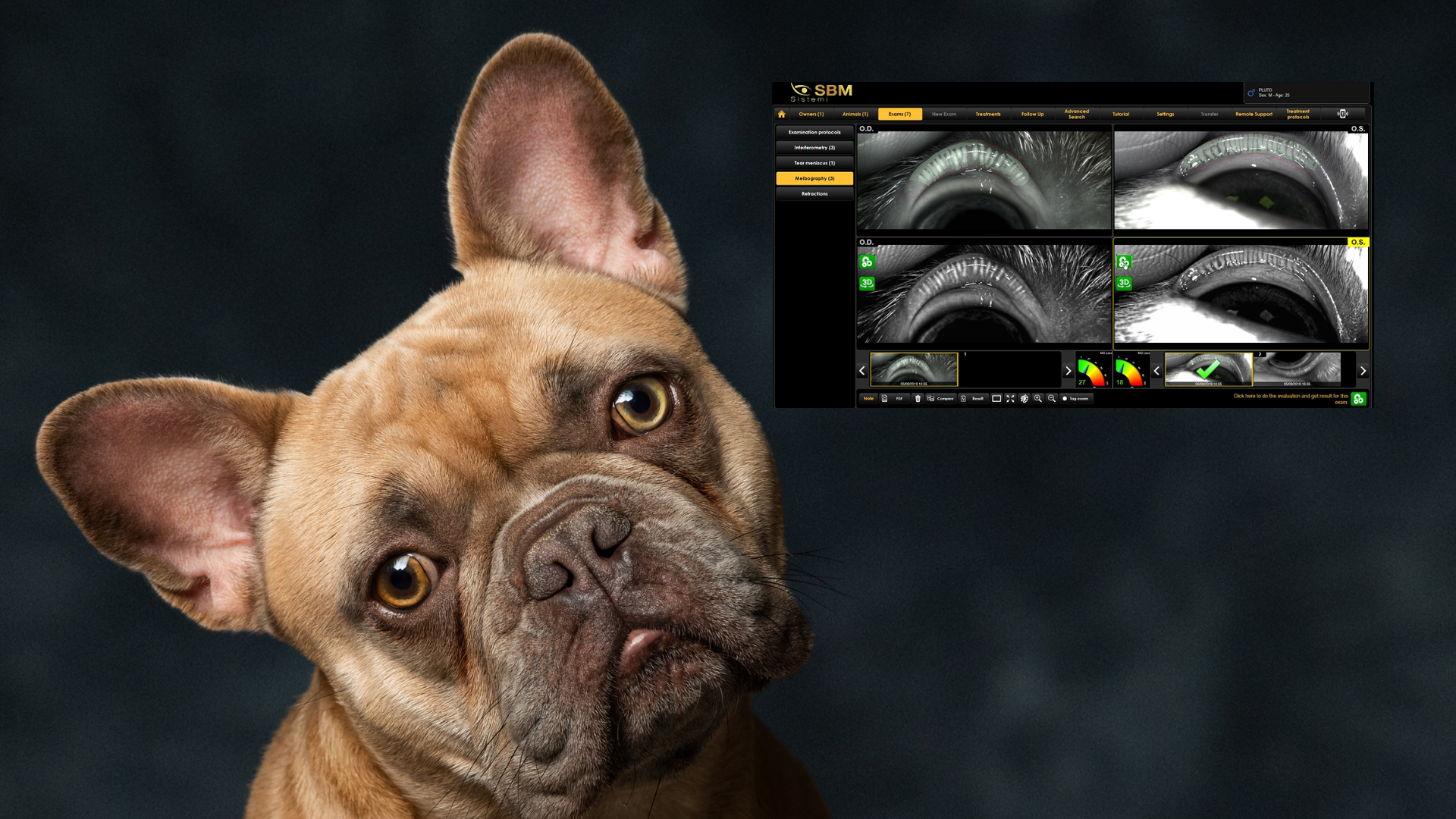
French Bulldogs have become one of the most popular dog breeds worldwide, but their unique facial anatomy also
predisposes them to ocular surface disorders. A recently published study in Frontiers in Veterinary Science
investigates tear film characteristics in healthy French Bulldogs and compares them with non-brachycephalic dogs,
using a comprehensive diagnostic approach that includes the OSA-Vet® system.
The results highlight qualitative differences in tear film integrity that could contribute to eye surface disease in
this breed and demonstrate the clinical value of objective tear film diagnostics like interferometry, NIBUT, and
meibography with OSA-Vet®.
Why Tear Film matters in dogs
The tear film plays a crucial role in maintaining ocular surface health, lubricating the eye, protecting against
infection, and ensuring clear vision. Disruptions in tear film quality or stability can lead to dry eye disease and
other surface disorders, especially in breeds with altered facial conformation like French Bulldogs.
Veterinarians distinguish between two main types of dry eye:
- Aqueous deficient dry eye (ADDE): insufficient tear production
- Evaporative dry eye (EDED): excessive tear evaporation due to a deficient lipid layer or film instability
This study focused on identifying qualitative tear film abnormalities, especially evaporative dry eye signs.
How the study was conducted
Researchers examined a group of healthy French Bulldogs alongside healthy dogs of mesocephalic and dolichocephalic breeds (non-brachycephalic controls). All dogs underwent:
- Full ophthalmic exam
- Tear film osmolarity measurement
- Comprehensive tear film analysis with OSA-Vet®, including:
- Interferometry (lipid layer assessment)
- Non-invasive tear break-up time (NIBUT)
- Tear meniscus height (TMH)
- Meibography (Meibomian gland evaluation)
Tear osmolarity was also measured with a separate device to explore whether this could help differentiate dry eye types.
Key findings
The study revealed several consistent differences between French Bulldogs and non-brachycephalic dogs:
- Shorter Tear Film Stability (NIBUT)
French Bulldogs had significantly lower NIBUT values, indicating a tear film that breaks up earlier after blinking, a hallmark of evaporative dry eye disease. - Lower Interferometry Scores
Interferometry with OSA-Vet® showed reduced lipid layer quality in French Bulldogs, meaning the protective oily layer was thinner or less stable, another sign pointing toward evaporative dry eye. - Higher Tear Meniscus Height
Although these dogs didn’t produce less tear volume, the higher tear meniscus height suggests reflex tearing, likely a response to rapid tear evaporation and eye surface irritation. - Similar Tear Osmolarity
Contrary to some expectations, tear osmolarity did not differ significantly between groups, indicating that tear chemistry alone may not distinguish dry eye types in this context. - Meibomian Gland Differences
Meibography also suggested greater Meibomian gland changes in French Bulldogs, potentially contributing to the instability of the lipid layer.
What this means for vets and pet owners
The study’s findings show that:
- Healthy French Bulldogs often show signs of evaporative dry eye disease (EDED) even without overt clinicalcomplaints.
- Standard tear volume tests may not be enough to detect subtle tear film dysfunction in brachycephalic dogs.
- Advanced diagnostics like OSA-Vet® interferometry, NIBUT, and meibography add significant value for earlydetection and tailored management of tear film disorders.
These insights can help veterinarians identify tear film deficiencies earlier and optimize treatment plans before secondary ocular surface disease develops.
Clinical implications and future perspectives
The findings of this study contribute to a better understanding of tear film dynamics in French Bulldogs and
highlight how breed-related anatomical features can influence ocular surface health, even in dogs considered
clinically healthy.
The results suggest that tear film instability and lipid layer alterations may be present before the onset of overt
clinical signs, reinforcing the importance of a qualitative assessment of the tear film in brachycephalic breeds. In
this context, diagnostic approaches that evaluate tear film stability, lipid layer characteristics, and Meibomian
gland morphology provide additional information compared to tear quantity measurements alone.
From a clinical perspective, these insights may support earlier identification of dogs at risk of developing ocular
surface disease and encourage more targeted monitoring strategies in predisposed breeds.
This study published in Frontiers in Veterinary Science offers a comprehensive analysis of tear film characteristics
in French Bulldogs compared to non-brachycephalic dogs.
These findings underline the value of a multimodal diagnostic approach to tear film evaluation and contribute to a
more nuanced understanding of ocular surface health in brachycephalic dogs. Further research will be useful to
clarify the clinical relevance of these alterations over time and their role in the development of ocular surface
disease.
This article is based on the peer-reviewed publication: Tear film characteristics in French Bulldogs compared to non-brachycephalic dogs (Frontiers in Veterinary Science, 2025).
A downloadable PDF of the original publication is attached here.
Read more
Objective vs subjective meibography: why quantification matters
Dry Eye in dogs and cats: beyond the Schirmer Test
The business case for Dry Eye clinics: revenue opportunities and patient retention
How corneal topography helps detect keratoconus in early stages
Automated Dry Eye diagnosis: how technology improves repeatability and workflow
Corneal Topography and Dry Eye: why both matter before cataract surgery
Tear film layers explained: lipid, aqueous and mucin
How to build a complete Dry Eye diagnostic workflow in your clinic
How to choose a Dry Eye diagnostic device for your practice
Meibomian gland dropout: early detection with advanced imaging
How objective Dry Eye diagnostics increase clinic revenue
Objective Optical Quality Metrics in Modern Ophthalmology
Regulatory Considerations When Importing CE Certified Ophthalmic Devices
Quantitative meibomian gland analysis: from images to objective data
OEM Opportunities in Ophthalmic Diagnostic Equipment
Best non-invasive tests for Dry Eye diagnosis in 2026
Transillumination meibography: clinical applications and advantages
What is a Tear Film Analyzer Device and how does it improve Dry Eye diagnosis?
Ocular surface assessment in high-volume clinics: efficiency without compromising accuracy
Beyond dry eye: other ocular surface disorders that impact visual quality
The role of ocular surface inflammation in post-surgical patient dissatisfaction
Ocular surface evaluation in contact lens wearers: what clinicians often miss
Environmental and lifestyle factors that alter tear film stability
Pre-surgical dry eye screening: which parameters really influence outcomes
Optimizing the ocular surface before cataract surgery to improve surgical accuracy and patient outcomes
Dry eye and cataract surgery: managing expectations through objective preoperative evaluation
Evidence-based guidelines for cataract surgery: improving outcomes through standardized clinical practice
Meibomian gland dysfunction: why early detection changes long-term patient outcomes
Meibography and MGD: from gland imaging to clinical decision making
Non-invasive tear film assessment: clinical advantages over fluorescein-based methods
Tear film analysis explained: LLT, NIBUT and tear meniscus in clinical practice
Why traditional dry eye tests are not enough: limits of Schirmer and TBUT in modern practice
Dry eye diagnosis today: why ocular surface analysis is no longer optional

Attention: The information on this page is intended exclusively for qualified healthcare professionals.


