Smart Studio
Smart Studio

Lens Effect 3D
Lens Effect 3D

Optic Studio
Optic Studio
Tear film analysis explained: LLT, NIBUT and tear meniscus in clinical practice
January 15, 2026
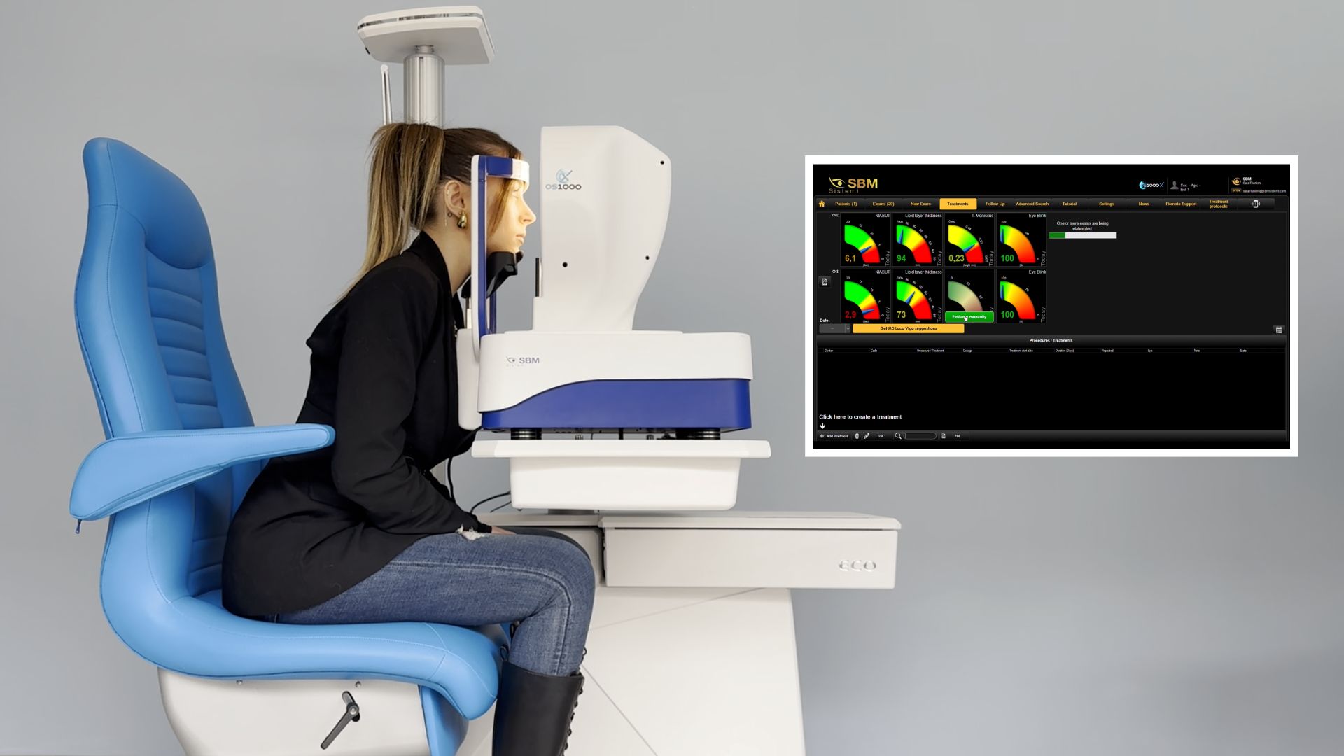
Dry Eye Disease (DED) is a multifactorial condition that cannot be fully understood through symptoms alone. Tear film
instability, excessive evaporation, reduced tear volume, and ocular surface alterations often coexist, making
objective tear film analysis an essential part of modern clinical practice.
Among the most relevant non-invasive parameters used today are Lipid Layer Thickness (LLT), Non-Invasive Break-Up
Time (NIBUT), and tear meniscus evaluation, commonly expressed as Tear Meniscus Height (TMH). Together, these
measurements provide a clear and practical overview of tear film quality and behavior.
The clinical value of tear film analysis
Patient-reported symptoms frequently show poor correlation with clinical signs. Objective tear film measurements help
clinicians overcome this limitation by offering repeatable data that support diagnosis, treatment planning, and
follow-up.
In everyday practice, tear film analysis allows clinicians to:
- Identify the main dry eye mechanism (evaporative, aqueous-deficient, or mixed)
- Establish a reliable baseline before treatment
- Monitor therapeutic effectiveness over time
- Improve patient understanding using visual outputs
Non-contact diagnostic methods are particularly advantageous because they are fast, comfortable for patients, and suitable for routine examinations.
LLT: Lipid Layer Thickness and evaporation control
The lipid layer plays a crucial role in reducing tear evaporation. LLT is typically assessed through interferometric
imaging, which visualizes lipid layer thickness and distribution across the tear film.
Clinically, LLT assessment is useful to:
- Detect evaporative dry eye
- Support the diagnosis of Meibomian Gland Dysfunction
- Guide lid-focused therapies
- Monitor response to treatments targeting the lipid layer
Because LLT is measured non-invasively and produces intuitive visual data, it also enhances patient education and treatment compliance.
NIBUT: Non-Invasive Break-Up Time and tear film stability
NIBUT measures the time interval between a blink and the first disruption of the tear film without using fluorescein
dye. This approach avoids reflex tearing and provides a more natural representation of tear film behavior.
In clinical practice, NIBUT helps to:
- Quantify tear film stability
- Identify early signs of dry eye
- Evaluate blinking quality and surface wetting
- Distinguish localized from diffuse instability
Advanced systems may also provide break-up maps and dynamic graphs, helping clinicians visualize how tear film instability develops over time.
Tear meniscus and Tear Meniscus Height
The tear meniscus is the reservoir of tears located along the eyelid margin. Measuring its height and regularity
offers a practical estimate of tear volume and distribution.
Tear meniscus evaluation supports:
- Identification of aqueous-deficient dry eye
- Assessment of tear availability at the ocular surface
- Treatment decisions involving tear supplementation or conservation
- Follow-up evaluation after therapeutic interventions
While TMH alone does not define a diagnosis, it becomes highly informative when combined with LLT and NIBUT.
Integrating LLT, NIBUT and tear meniscus in diagnosis
Using these three parameters together enables a simple and effective diagnostic framework:
- Reduced LLT indicates increased evaporation
- Reduced NIBUT confirms tear film instability
- Reduced tear meniscus suggests limited tear volume
Common clinical patterns include:
- Low LLT with low NIBUT and normal tear meniscus, typical of evaporative dry eye
- Normal LLT with low NIBUT and low tear meniscus, suggesting aqueous-deficient or mixed dry eye
- Reduced values across all parameters, indicating mixed dry eye
- Normal LLT and tear meniscus with reduced NIBUT, often linked to blinking or surface wetting issues
This integrated approach supports faster classification and more targeted treatment strategies.
A practical workflow for tear film evaluation
A streamlined tear film analysis workflow may include:
- Patient history and symptom assessment
- Non-invasive measurements of LLT, NIBUT, and tear meniscus
- Mechanism-based dry eye classification
- Patient education using visual reports
- Follow-up with repeat measurements to monitor improvement
Standardized protocols improve consistency and make follow-up comparisons more meaningful.
LLT, NIBUT, and tear meniscus assessment provide a reliable, non-invasive foundation for tear film evaluation in
clinical practice. When used together, these parameters help clinicians move beyond symptom-based management toward
targeted, physiology-driven dry eye care.
Integrating tear film analysis into routine examinations improves diagnostic accuracy, enhances patient
communication, and supports long-term monitoring, ultimately leading to better outcomes for patients with dry eye
disease.
Read more
Corneal Topography vs. Corneal Tomography: What Is the Difference?
When Dry Eye Treatment Isn't Working: A Diagnostic Approach
Demodex and Ocular Surface Disease: What Clinicians Should Know
Blepharitis and Dry Eye Understanding the Clinical Connection
Why Tear-Film Stability Matters for Visual Quality and Preoperative Measurements
How to Build a Comprehensive Dry Eye Diagnostic Workflow
Can MGD Be Detected Before Symptoms Appear?
Meibomian Gland Tortuosity, Shortening and Atrophy: What Do These Findings Mean?
Meibomian Gland Morphology: What Should Clinicians Look for in an Image?
The Role of Blinking in Tear Film Stability
Why Dry Eye Symptoms and Clinical Signs Don't Always Match
Evaporative vs. Aqueous-Deficient Dry Eye: How to Tell Them Apart
Dry Eye vs. Meibomian Gland Dysfunction: What Is the Difference?
Objective vs subjective meibography: why quantification matters
Dry Eye in dogs and cats: beyond the Schirmer Test
The business case for Dry Eye clinics: revenue opportunities and patient retention
How corneal topography helps detect keratoconus in early stages
Automated Dry Eye diagnosis: how technology improves repeatability and workflow
Corneal Topography and Dry Eye: why both matter before cataract surgery
Tear film layers explained: lipid, aqueous and mucin
How to build a complete Dry Eye diagnostic workflow in your clinic
How to choose a Dry Eye diagnostic device for your practice
Meibomian gland dropout: early detection with advanced imaging
How objective Dry Eye diagnostics increase clinic revenue
Objective Optical Quality Metrics in Modern Ophthalmology
Regulatory Considerations When Importing CE Certified Ophthalmic Devices
Quantitative meibomian gland analysis: from images to objective data
OEM Opportunities in Ophthalmic Diagnostic Equipment
Best non-invasive tests for Dry Eye diagnosis in 2026
Transillumination meibography: clinical applications and advantages
What is a Tear Film Analyzer Device and how does it improve Dry Eye diagnosis?
Ocular surface assessment in high-volume clinics: efficiency without compromising accuracy
Beyond dry eye: other ocular surface disorders that impact visual quality
The role of ocular surface inflammation in post-surgical patient dissatisfaction
Ocular surface evaluation in contact lens wearers: what clinicians often miss
Environmental and lifestyle factors that alter tear film stability
Tear Film health in French Bulldogs: what the latest study using OSA-Vet® reveals
Pre-surgical dry eye screening: which parameters really influence outcomes
Optimizing the ocular surface before cataract surgery to improve surgical accuracy and patient outcomes
Dry eye and cataract surgery: managing expectations through objective preoperative evaluation
Evidence-based guidelines for cataract surgery: improving outcomes through standardized clinical practice
Meibomian gland dysfunction: why early detection changes long-term patient outcomes
Meibography and MGD: from gland imaging to clinical decision making
Non-invasive tear film assessment: clinical advantages over fluorescein-based methods
Why traditional dry eye tests are not enough: limits of Schirmer and TBUT in modern practice
Dry eye diagnosis today: why ocular surface analysis is no longer optional

Attention: The information on this page is intended exclusively for qualified healthcare professionals.