


Smart Studio
Smart Studio

Lens Effect 3D
Lens Effect 3D

Optic Studio
Optic Studio
Objective Optical Quality Metrics in Modern Ophthalmology
April 15, 2026
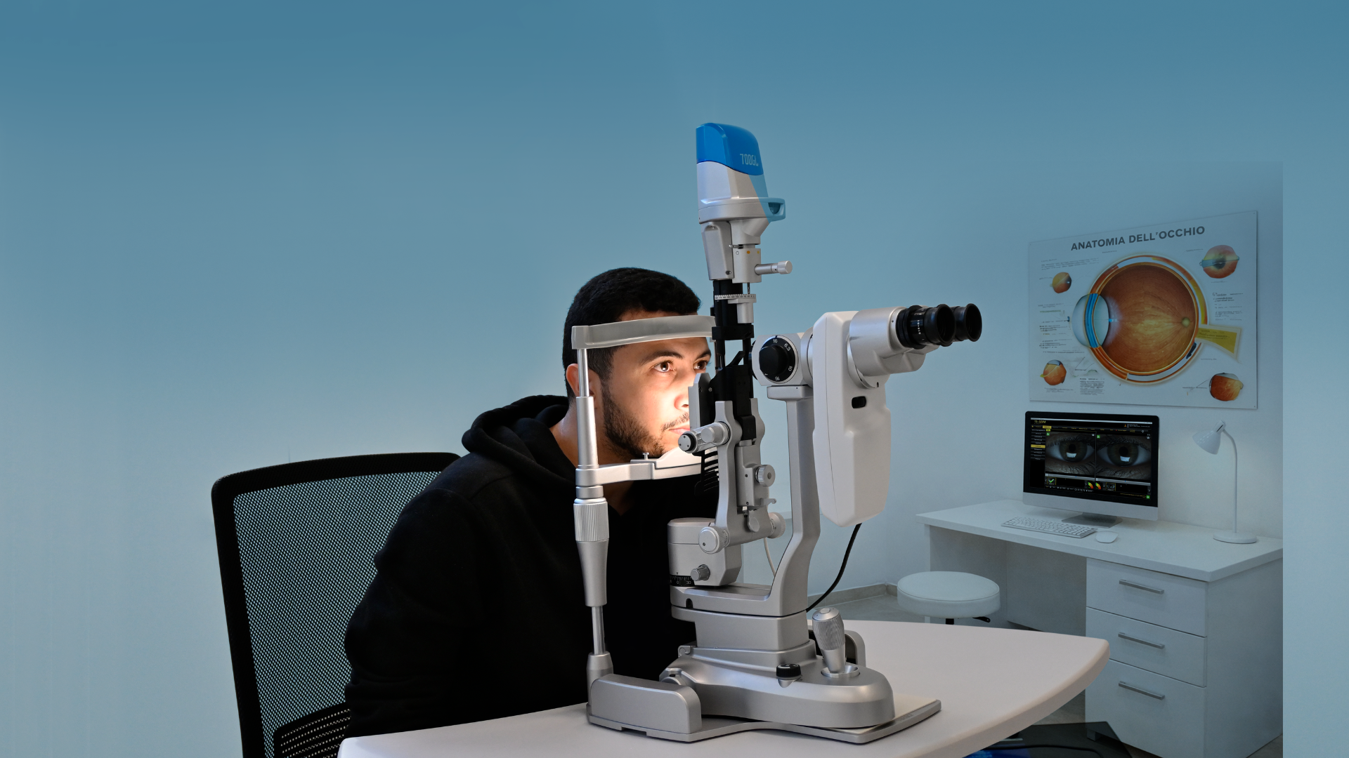
In modern ophthalmology, evaluating visual quality goes beyond standard visual acuity. Clinicians are increasingly
adopting objective technologies to better understand how patients truly see, especially in complex conditions
involving the ocular surface and tear film.
An optical quality diagnostic device allows eye care professionals to measure visual performance in a quantitative
and repeatable way, supporting more accurate diagnosis and more personalized treatment strategies.
Understanding optical quality metrics
Optical quality metrics describe how light is transmitted through the eye and how clearly it is focused on the
retina. Unlike subjective tests, these measurements provide objective data that can be tracked over time.
Key parameters include:
- Point Spread Function (PSF)
- Modulation Transfer Function (MTF)
- Strehl ratio
- Optical Scatter Index (OSI)
These metrics are particularly useful when visual complaints are not fully explained by refractive error alone.
The clinical value of an optical quality diagnostic device
An optical quality diagnostic device helps clinicians detect subtle alterations in vision that traditional exams may
overlook. This is especially important in patients with fluctuating vision, early cataract, or ocular surface
disorders.
In daily practice, these devices support:
- pre- and post-surgical evaluation
- assessment of intraocular scatter
- analysis of visual quality under real conditions
- monitoring of tear film stability
As highlighted in SBM’s approach to diagnostics, modern ophthalmology is moving toward integrated, data-driven evaluation rather than isolated tests.
Integration with dry eye diagnostics
One of the most relevant applications of optical quality metrics is in dry eye disease. Tear film instability
directly affects optical performance, often causing visual fluctuations that patients struggle to describe.
This is why optical quality assessment is increasingly combined with tear film analysis workflows.
You can explore this approach further in SBM’s article on tear film technology:
→ What is a tear film analyzer device
By combining optical data with tear film evaluation, clinicians can better understand the link between symptoms and
ocular surface alterations.
Supporting ocular surface analysis
Optical quality diagnostics are also closely connected to broader ocular surface assessment. Meibomian gland
dysfunction, lipid layer alterations, and blink dynamics all influence visual clarity.
SBM content consistently highlights the importance of a multi-parametric approach to dry eye and ocular surface
disease, where different diagnostic tools work together.
Related insights can be found in:
→ Non-invasive dry eye tests
→ Meibography in clinical practice
This integrated model allows clinicians to:
- correlate structural and functional data
- improve diagnostic accuracy
- monitor treatment outcomes more effectively
Why objective metrics are becoming essential
The shift toward objective diagnostics reflects a broader trend in ophthalmology: reducing variability and improving
reproducibility.
Using an optical quality diagnostic device provides:
- objective, repeatable measurements
- better understanding of patient-reported symptoms
- improved communication through visual data
- stronger clinical decision-making
For clinics specializing in dry eye and ocular surface disorders, this represents a significant upgrade in diagnostic capability.
Choosing the right diagnostic approach
When selecting technologies for a modern ophthalmic practice, it is important to consider how well different tools
work together.
Rather than using standalone devices, many clinics are adopting integrated platforms that combine:
- optical quality analysis
- tear film evaluation
- meibography
- non-invasive diagnostics
SBM Sistemi’s diagnostic philosophy reflects this trend, focusing on comprehensive dry eye platforms that streamline workflow while maintaining high diagnostic precision.
Objective optical quality metrics are redefining how clinicians assess vision. By using an optical quality diagnostic
device, ophthalmologists can move beyond subjective evaluation and gain deeper insight into visual performance.
When combined with tear film analysis and ocular surface diagnostics, these technologies enable a more complete
understanding of dry eye disease and related conditions.
As ophthalmology continues to evolve, integrating objective optical quality assessment into daily practice is
becoming essential for delivering accurate, data-driven patient care.
Read more
Objective vs subjective meibography: why quantification matters
Dry Eye in dogs and cats: beyond the Schirmer Test
The business case for Dry Eye clinics: revenue opportunities and patient retention
How corneal topography helps detect keratoconus in early stages
Automated Dry Eye diagnosis: how technology improves repeatability and workflow
Corneal Topography and Dry Eye: why both matter before cataract surgery
Tear film layers explained: lipid, aqueous and mucin
How to build a complete Dry Eye diagnostic workflow in your clinic
How to choose a Dry Eye diagnostic device for your practice
Meibomian gland dropout: early detection with advanced imaging
How objective Dry Eye diagnostics increase clinic revenue
Regulatory Considerations When Importing CE Certified Ophthalmic Devices
Quantitative meibomian gland analysis: from images to objective data
OEM Opportunities in Ophthalmic Diagnostic Equipment
Best non-invasive tests for Dry Eye diagnosis in 2026
Transillumination meibography: clinical applications and advantages
What is a Tear Film Analyzer Device and how does it improve Dry Eye diagnosis?
Ocular surface assessment in high-volume clinics: efficiency without compromising accuracy
Beyond dry eye: other ocular surface disorders that impact visual quality
The role of ocular surface inflammation in post-surgical patient dissatisfaction
Ocular surface evaluation in contact lens wearers: what clinicians often miss
Environmental and lifestyle factors that alter tear film stability
Tear Film health in French Bulldogs: what the latest study using OSA-Vet® reveals
Pre-surgical dry eye screening: which parameters really influence outcomes
Optimizing the ocular surface before cataract surgery to improve surgical accuracy and patient outcomes
Dry eye and cataract surgery: managing expectations through objective preoperative evaluation
Evidence-based guidelines for cataract surgery: improving outcomes through standardized clinical practice
Meibomian gland dysfunction: why early detection changes long-term patient outcomes
Meibography and MGD: from gland imaging to clinical decision making
Non-invasive tear film assessment: clinical advantages over fluorescein-based methods
Tear film analysis explained: LLT, NIBUT and tear meniscus in clinical practice
Why traditional dry eye tests are not enough: limits of Schirmer and TBUT in modern practice
Dry eye diagnosis today: why ocular surface analysis is no longer optional

Attention: The information on this page is intended exclusively for qualified healthcare professionals.


