


Smart Studio
Smart Studio

Lens Effect 3D
Lens Effect 3D

Optic Studio
Optic Studio
Meibomian Gland Tortuosity, Shortening and Atrophy: What Do These Findings Mean?
October 19, 2026
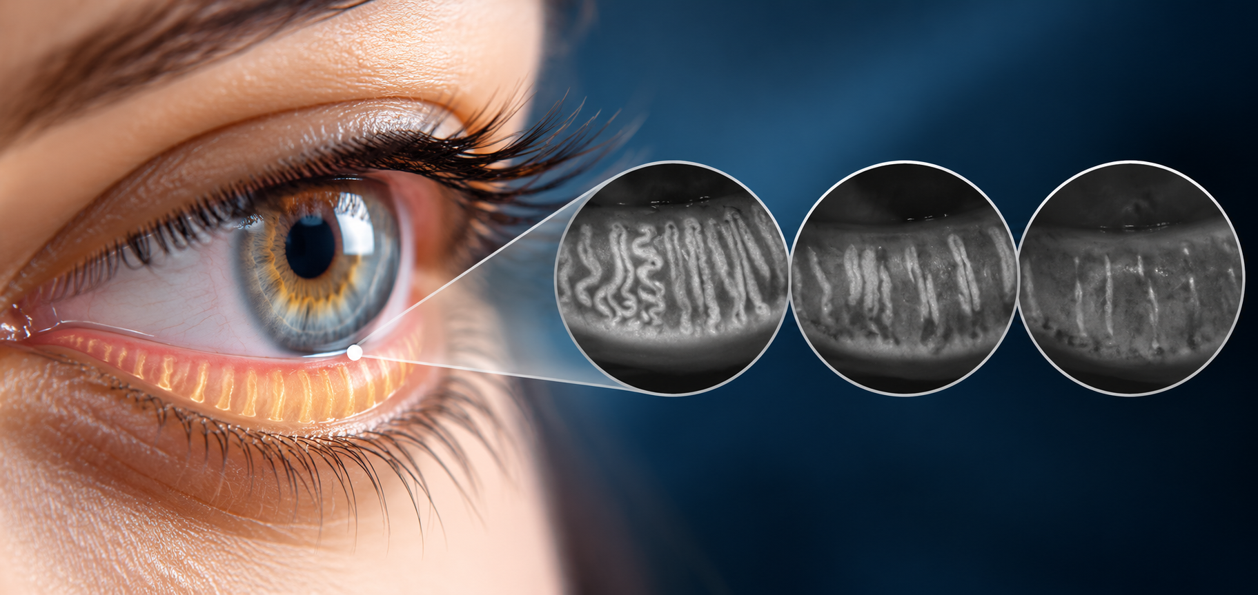
Meibography has made it possible to visualize the meibomian glands in much greater detail than was possible with
routine clinical examination alone. When reviewing a meibography image, clinicians often focus first on gland
dropout. However, the morphology of the glands can provide additional information. Changes such as tortuosity,
shortening, irregularity and atrophy may appear before extensive gland loss becomes evident. Understanding these
findings and, importantly, understanding their limitations is essential for interpreting meibography correctly.
A meibography image should not simply be classified as "normal" or "abnormal." Instead, clinicians can evaluate the
overall gland architecture and consider how structural findings relate to gland function, tear-film stability and
ocular-surface symptoms. This approach can help transform meibography from a simple imaging technique into a more
informative component of ocular-surface assessment.
Understanding Normal Meibomian Gland Anatomy
The meibomian glands are specialized sebaceous glands embedded within the tarsal plates of the eyelids.
They consist broadly of:
- A central duct
- Secretory acini
- Ductules
- A terminal opening at the lid margin
Their secretion, meibum, contributes to the lipid layer of the tear film. Although the general architecture is consistent, the appearance of individual glands can vary considerably between patients and between different regions of the eyelids. This anatomical variability is important. Not every curved or irregular gland should be interpreted as pathological. Clinical interpretation should therefore focus on patterns of change, rather than isolated morphological features.
What Is Gland Tortuosity?
Tortuosity refers to curvature or twisting of the gland structure. Some degree of curvature may occur naturally.
However, increased tortuosity or irregular gland architecture can become more apparent in patients with meibomian
gland dysfunction. When evaluating tortuosity, clinicians can consider how pronounced the curvature is, whether
multiple glands show the same pattern or if changes are localized or diffuse. Another observation can be whether
tortuosity is associated with shortening or if functional abnormalities are also present. The presence of tortuosity
alone should therefore not automatically lead to a diagnosis of MGD.
The precise mechanisms behind changes in gland morphology are complex. Potential contributing factors include
chronic gland dysfunction with duct obstruction and/or altered meibum flow; tissue remodeling; inflammation and
age-related changes.
These processes may influence the architecture of the gland over time. However, morphology should always be
interpreted within the broader clinical picture.
What Is Gland Shortening?
Gland shortening occurs when a gland appears reduced in length compared with the expected anatomical pattern. It can
be subtle. Instead of a complete, elongated gland extending through much of the tarsal plate, the visible gland may
appear truncated.
Shortening can occur together with:
- Tortuosity
- Gland distortion
- Partial dropout
- Reduced gland density
The significance of shortening depends on its extent, distribution and association with other findings. Additionally, it is important to distinguish between gland shortening and gland dropout. These two findings are related but not identical.
- Gland shortening means that a gland is still visible but appears reduced in length.
- Gland dropout refers to loss or absence of visible gland tissue.
A patient may therefore show significant gland shortening before complete gland loss becomes apparent. This distinction can be useful when monitoring structural changes over time. A gland that becomes progressively shorter may represent a different stage of morphological change from a gland that is no longer visible.
What Is Meibomian Gland Atrophy?
Atrophy generally refers to a reduction or degeneration of glandular tissue. On meibography, this may be reflected by
changes such as reduced gland size, shortened glands, irregular gland architecture or areas of gland loss.
The term "atrophy" should be used carefully because the image provides structural information, while the underlying
biological process may be more complex. For this reason, describing the actual observed morphology can sometimes be
more informative than applying a broad label.
If glandular structure becomes compromised, secretion may also be affected. This can contribute to changes in the
tear-film lipid layer and potentially increase evaporation.
However structural changes do not always produce the same degree of functional impairment. This is one of the most
important principles in meibography interpretation. A patient may have visible gland abnormalities but relatively
preserved gland function. Another patient may have significant functional MGD with comparatively limited structural
changes. This distinction explains why meibography should be combined with functional assessment. The combination of
structural and functional information is generally more informative than either alone.
The Importance of Gland Distribution
Morphological changes should also be evaluated according to their distribution. Patterns may provide useful context.
For example, localized abnormalities may have a different clinical interpretation from widespread gland changes.
The upper and lower eyelids have different anatomical characteristics. The upper eyelid generally contains more
meibomian glands and longer gland structures. As a result, direct numerical comparisons between upper and lower
eyelids should be interpreted carefully. For longitudinal monitoring, it is preferable to compare measurements
obtained from the same anatomical region whenever possible.
Additionally, Meibomian Gland morphology can differ between the two eyes. Some degree of asymmetry is possible.
However, pronounced differences may be clinically relevant, particularly when structural asymmetry corresponds to
differences in symptoms or functional measurements. A bilateral comparison can therefore provide additional context
during examination.
Finally, changes in gland structure and function may occur as part of the aging process. This means that
morphological findings should always be interpreted in relation to the patient's age and clinical context. An
appearance that may be unusual in a young patient may not have the same significance in an older patient. Age should
therefore be considered when evaluating quantitative parameters and comparing patients.
Why a Single Morphological Finding Is Not Enough
Imagine a patient with slightly tortuous glands but:
- Normal meibum
- Good gland expressibility
- Stable tear film
- No significant symptoms
It would be inappropriate to conclude that the patient necessarily has clinically significant MGD based solely on gland tortuosity. Now consider a different patient with:
- Marked gland shortening and significant gland dropout
- Thickened meibum and poor expressibility
- Short tear-film breakup time
- Ocular discomfort
The combined picture is much more compelling. This illustrates why morphology should be interpreted as part of a
multidimensional assessment.
Traditionally, clinicians have described meibography findings using qualitative terms such as mild, moderate and
severe. The human eye is excellent at recognizing patterns, but visual grading can be influenced by observer
experience. Objective imaging provides a permanent record that can be reviewed again. The objective is not to
eliminate clinical interpretation. Instead, quantitative analysis can complement the clinician's assessment with
measurable parameters.
Why Gland Length Can Be Useful
Gland length is one example of a parameter that can be monitored over time. If an individual gland or a region of
glands becomes progressively shorter, quantitative documentation may make this change easier to identify. However,
measurements must be standardized. Changes in eyelid eversion, image positioning and quality and region analyzed can
influence apparent gland length. Therefore, reproducibility is essential.
A major advantage of imaging is the ability to create a baseline. Suppose a patient undergoes meibography at their
first examination. The images document the current morphology. At subsequent visits, the clinician can compare the
new images with the baseline.
If follow-up images are acquired differently from baseline images, apparent differences may be caused by technique
rather than biology. For this reason, standardized imaging should follow a consistent acquisition process, so that
comparisons can really be meaningful.
Morphology and Treatment Monitoring
Meibography can also be useful when evaluating patients over the course of treatment. However, clinicians should be cautious about expecting structural recovery simply because symptoms improve. Symptoms and function may change however gland anatomy is rarely improving. This does not necessarily indicate treatment failure. Structural tissue loss and functional improvement are different outcomes. Therefore, treatment monitoring should consider both short-term functional changes and longer-term structural observations.
Can Glands Regrow?
One of the questions patients often ask is whether lost meibomian glands can return. At present, complete restoration
of gland tissue after established gland dropout should not be assumed. This makes prevention of progressive gland
damage and early identification of structural abnormalities important areas of clinical attention. However,
improvement in gland function does not necessarily require anatomical restoration of every lost gland. Residual
glands may continue to contribute to meibum production even when some gland tissue has been lost.
This distinction is important when communicating with patients.
How Should Clinicians Report Morphological Findings?
A useful report should ideally be descriptive and clinically meaningful. The findings can be correlated with
functional and symptomatic information. This makes the report more useful for future follow-up.
When evaluating gland morphology, consider the following sequence.
- Image quality: Can the glands be clearly visualized?
- Anatomical region: Which eyelid and which region are being assessed?
- Gland density: Are the glands regularly distributed?
- Gland length: Are glands full length or shortened?
- Gland shape: Are they straight, curved or irregular?
- Tortuosity: Is curvature within expected anatomical variation or clearly pronounced?
- Gland dropout: Are there areas of partial or complete gland loss?
- Symmetry: Are the findings similar between the two eyes?
- Functional correlation: Do the morphological findings correspond with meibum or expressibility abnormalities?
- Tear-film correlation: Is there evidence of tear-film instability or increased evaporation?
This structured approach can improve consistency and reduce the risk of overinterpreting individual findings.
Conclusion
Meibomian gland morphology provides valuable information about the structural condition of the ocular surface. While
gland dropout remains one of the most recognized findings on meibography, other features, including tortuosity,
shortening, density and architectural changes, can provide additional information.
The challenge is to interpret these findings correctly.
Not every morphological variation represents disease, and structural abnormalities do not always correspond directly
to symptoms or functional impairment. For this reason, the most informative approach combines meibography with
functional assessment, tear-film analysis and clinical examination. Quantitative measurements can further support
this process by transforming visual observations into measurable parameters that can be documented and compared over
time.
As ocular-surface diagnostics continue to evolve, the combination of high-quality imaging, quantitative analysis and
clinical expertise has the potential to make meibomian gland assessment increasingly objective, reproducible and
useful for longitudinal patient care.
Key Takeaways
- Meibomian gland morphology includes more than gland dropout.
- Tortuosity, shortening and architectural changes can provide additional information.
- Gland shortening and gland dropout are related but distinct findings.
- Some morphological variation is normal and should not automatically be interpreted as disease.
- Structural abnormalities do not necessarily correspond directly to functional impairment.
- MGD can occur with relatively preserved gland structure.
- Significant gland loss can occur with relatively mild symptoms.
- Meibography should be interpreted alongside functional and symptomatic information.
- Quantitative measurements can complement qualitative assessment.
- Standardized imaging is essential for meaningful comparison in the long term.
- Structural changes may evolve differently from symptoms and functional parameters.
- The goal of meibography is not simply to identify gland loss, but to understand gland structure within the broader context of ocular-surface health.
Frequently Asked Questions
What does meibomian gland tortuosity mean?
Tortuosity describes curvature or twisting of the meibomian glands. Some curvature can be part of normal anatomical variation, so tortuosity should be interpreted together with other morphological and functional findings.
Is gland shortening the same as gland dropout?
No. A shortened gland remains partially visible, whereas dropout refers to the absence or loss of visible gland tissue.
What is meibomian gland atrophy?
Atrophy generally refers to a reduction or degeneration of glandular tissue. On meibography, this may appear as reduced gland size, shortening, decreased density or gland loss.
Can you have MGD without gland dropout?
Yes. Functional MGD can occur even when the glands appear relatively well structured.
Can gland dropout occur without severe symptoms?
Yes. Structural gland loss and symptom severity do not always correlate directly.
Can meibomian glands grow back after dropout?
Established gland dropout should not be assumed to be completely reversible. However, gland function and symptoms can potentially change independently of structural gland loss.
Why is quantitative meibography useful?
Quantitative analysis can provide measurable information about gland morphology and support more objective comparisons between examinations.
Should meibography be performed on both eyes?
When clinically appropriate, bilateral imaging can help identify asymmetry and provide a more complete assessment.
Read more
Meibomian Gland Morphology: What Should Clinicians Look for in an Image?
The Role of Blinking in Tear Film Stability
Why Dry Eye Symptoms and Clinical Signs Don't Always Match
Evaporative vs. Aqueous-Deficient Dry Eye: How to Tell Them Apart
Dry Eye vs. Meibomian Gland Dysfunction: What Is the Difference?
Objective vs subjective meibography: why quantification matters
Dry Eye in dogs and cats: beyond the Schirmer Test
The business case for Dry Eye clinics: revenue opportunities and patient retention
How corneal topography helps detect keratoconus in early stages
Automated Dry Eye diagnosis: how technology improves repeatability and workflow
Corneal Topography and Dry Eye: why both matter before cataract surgery
Tear film layers explained: lipid, aqueous and mucin
How to build a complete Dry Eye diagnostic workflow in your clinic
How to choose a Dry Eye diagnostic device for your practice
Meibomian gland dropout: early detection with advanced imaging
How objective Dry Eye diagnostics increase clinic revenue
Objective Optical Quality Metrics in Modern Ophthalmology
Regulatory Considerations When Importing CE Certified Ophthalmic Devices
Quantitative meibomian gland analysis: from images to objective data
OEM Opportunities in Ophthalmic Diagnostic Equipment
Best non-invasive tests for Dry Eye diagnosis in 2026
Transillumination meibography: clinical applications and advantages
What is a Tear Film Analyzer Device and how does it improve Dry Eye diagnosis?
Ocular surface assessment in high-volume clinics: efficiency without compromising accuracy
Beyond dry eye: other ocular surface disorders that impact visual quality
The role of ocular surface inflammation in post-surgical patient dissatisfaction
Ocular surface evaluation in contact lens wearers: what clinicians often miss
Environmental and lifestyle factors that alter tear film stability
Tear Film health in French Bulldogs: what the latest study using OSA-Vet® reveals
Pre-surgical dry eye screening: which parameters really influence outcomes
Optimizing the ocular surface before cataract surgery to improve surgical accuracy and patient outcomes
Dry eye and cataract surgery: managing expectations through objective preoperative evaluation
Evidence-based guidelines for cataract surgery: improving outcomes through standardized clinical practice
Meibomian gland dysfunction: why early detection changes long-term patient outcomes
Meibography and MGD: from gland imaging to clinical decision making
Non-invasive tear film assessment: clinical advantages over fluorescein-based methods
Tear film analysis explained: LLT, NIBUT and tear meniscus in clinical practice
Why traditional dry eye tests are not enough: limits of Schirmer and TBUT in modern practice
Dry eye diagnosis today: why ocular surface analysis is no longer optional

Attention: The information on this page is intended exclusively for qualified healthcare professionals.


