


Smart Studio
Smart Studio

Lens Effect 3D
Lens Effect 3D

Optic Studio
Optic Studio
Dry Eye vs. Meibomian Gland Dysfunction: What Is the Difference?
October 1, 2026
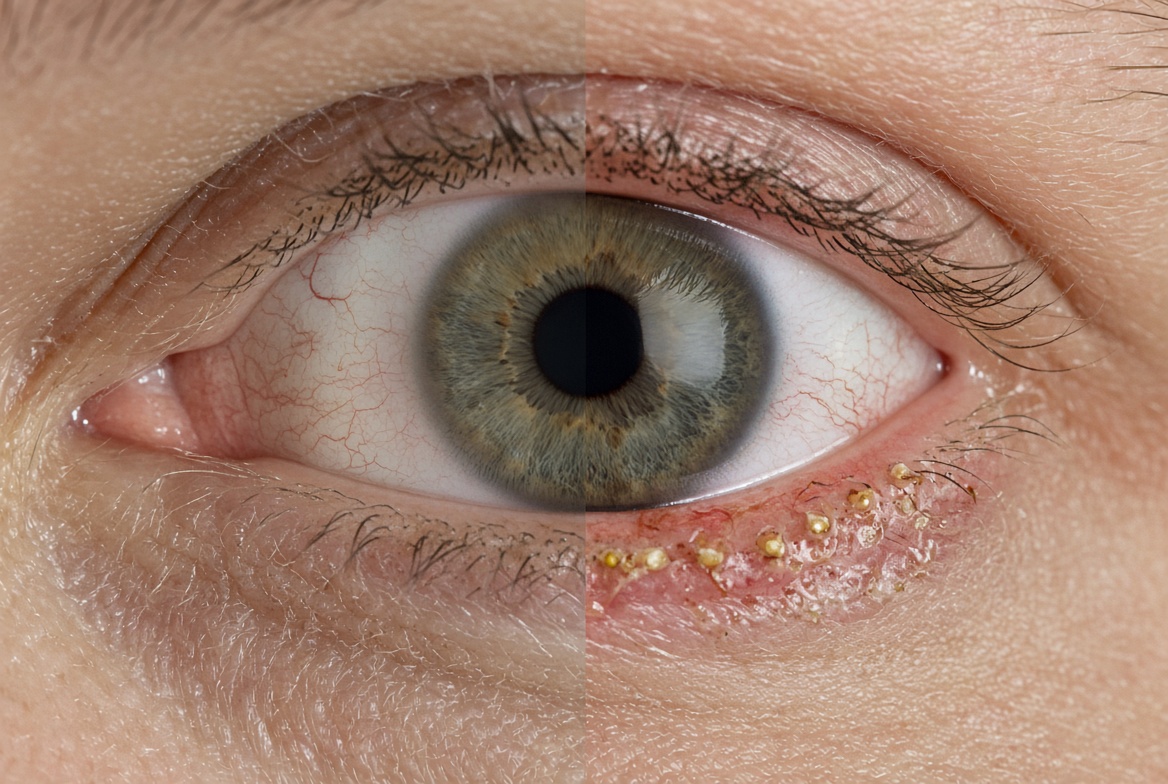
Dry Eye Disease (DED) and Meibomian Gland Dysfunction (MGD) are two of the most common ocular surface disorders
encountered in clinical practice. Although they are closely related, these two problems are not the same condition.
Understanding the difference between DED and MGD is important because patients may present with similar symptoms,
while the underlying mechanisms, and therefore the most appropriate diagnostic and therapeutic approach, can be
different.
MGD is one of the leading causes of evaporative dry eye, but not every patient with dry eye has clinically
significant MGD. Conversely, patients with early or asymptomatic DED may already show structural changes in the
meibomian glands before experiencing significant symptoms. For this reason, a comprehensive ocular surface
assessment should go beyond symptoms alone and consider both tear-film function and meibomian gland structure and
function.
What Is Dry Eye Disease?
Dry eye disease is a multifactorial disorder of the ocular surface characterized by a loss of tear-film homeostasis. It can involve abnormalities of the tears, ocular surface, eyelids, and sensory pathways. Even though the most recent studies have introduced a diverse range of subtype drivers to the pathology, in clinical practice, dry eye is often broadly divided into two major subtype classifications:
- Aqueous-deficient dry eye, in which the lacrimal glands do not produce sufficient aqueous tears.
- Evaporative dry eye, in which tear evaporation is excessive, often because of an abnormal tear-film lipid layer.
These mechanisms frequently overlap. A patient may therefore have both reduced tear production and increased
evaporation.
Typical symptoms include:
- Dryness, burning or stinging
- Foreign-body sensation
- Fluctuating vision
- Ocular fatigue and redness
- Light sensitivity
- Eccessive lacrimation
However, symptoms alone do not always reflect the severity of ocular surface disease. Indeed, one of the challenges
with DED and MGD is the imperfect relationship between symptoms and objective findings. Some patients with
significant structural or functional abnormalities may report relatively few symptoms, while others can experience
substantial discomfort despite relatively limited clinical signs. This is one of the reasons why objective
examination is increasingly important in dry-eye assessment.
Several factors can influence symptoms, including corneal nerve sensitivity, inflammation, environmental exposure,
contact lens wear, screen use, blink behavior, age, previous ocular surgery, systemic and medication-related
factors.
Therefore, asking patients whether their eyes feel dry is important, but it is only one part of the diagnostic
process. Objective measurements can provide additional information that symptoms cannot.
What Is Meibomian Gland Dysfunction?
Meibomian Gland Dysfunction is a chronic disorder of the meibomian glands characterized by terminal duct obstruction
and/or qualitative or quantitative changes in glandular secretion.
The meibomian glands are located within the tarsal plates of the eyelids. Their primary role is to produce meibum,
the lipid component of the tear film which helps reduce excessive tear evaporation and contributes to tear-film
stability.
When the glands become dysfunctional, several changes may occur. From altered meibum quality to increased secretion
viscosity; from duct obstruction, which leads to reduced gland secretion, to inflammation around the glands. These
problems can lead to gland shortening or atrophy, and in worse cases partial or complete gland dropouts. These
changes can compromise the lipid layer of the tear film and increase evaporation. The result can be an unstable tear
film and the development or worsening of evaporative dry eye.
The simplest way to understand the relationship is: MGD is a condition affecting the meibomian glands, while dry eye
disease is a broader disorder of tear-film and ocular-surface homeostasis. Specifically, MGD can cause or contribute
to dry eye, but dry eye can also result from other mechanisms. This distinction matters because treating the ocular
surface without identifying the underlying mechanism may result in incomplete or inefficient management.
Why MGD Is So Important in Dry Eye
The lipid layer of the tear film plays an important role in limiting evaporation. When meibomian gland secretion is
reduced or abnormal, the lipid layer may become insufficient or dysfunctional.
This can lead to increased tear evaporation, faster tear-film breakup, hyperosmolarity, inflammation and even
further gland dysfunction. The combination of these elements can create a self-reinforcing cycle. In other words,
MGD can contribute to tear-film instability, while chronic ocular-surface inflammation may in turn contribute to
further meibomian gland dysfunction.
For clinicians, identifying where a patient sits within this cycle can be more useful than simply assigning a
general diagnosis of "dry eye."
How Can MGD Be Identified?
A complete assessment of MGD should consider both function and structure.
Functional assessment
The clinician may subjectively assess meibum quality by evaluating the color, consistency, and viscosity of the expressed meibum. The ease of meibum expression may also be assessed to identify potential gland obstruction or dysfunction. Objective measurements of lipid layer thickness and tear-film stability, together with the assessment of symptoms during blinking or visually demanding tasks, may provide additional information. Collectively, these parameters can help characterize meibomian gland function and its contribution to tear-film stability.
Structural assessment
Meibography has become an important tool for evaluating the structure of the meibomian glands. It provides a way to
visualize the meibomian glands directly. Depending on the imaging system and examination protocol, clinicians can
assess characteristics such as: gland visibility, length, shape, tortuosity, shortening, loss area or complete gland
dropout.
Structural imaging is particularly valuable because gland abnormalities may exist even when symptoms are limited.
Unlike a conventional clinical examination, which may provide indirect information about gland function, meibography
allows the clinician to visualize gland morphology. This can help answer quantitative questions related to glands’
presence shape and structure.
A visual assessment of a meibography image can be useful, but it may also be subjective. Two observers may interpret
the same image differently, particularly when gland abnormalities are subtle. Conversely, quantitative analysis can
complement clinical judgment by transforming images into measurable parameters. This is particularly useful when the
goal is to monitor changes over time or compare findings between examinations.
Why Early Identification of MGD Matters
Meibomian gland dysfunction can be progressive. Once glandular tissue is lost, restoring the original anatomy may be difficult or impossible. For this reason, identifying structural changes at an early stage may be clinically valuable. This is especially relevant in patients with risk factors for ocular surface disease, including increasing age, chronic eyelid disease, contact lens wear, prolonged screen use and previous ocular surgery.
MGD Before Cataract or Refractive Surgery
The distinction between dry eye and MGD becomes particularly relevant before ocular surgery.
Patients undergoing cataract or refractive procedures may require precise preoperative measurements. An unstable
tear film can affect the quality and repeatability of some ocular measurements, potentially complicating surgical
planning. For this reason, evaluating and, when necessary, optimizing the ocular surface before final measurements
can be an important part of the preoperative workflow.
Identifying MGD is particularly useful because a patient may have relatively mild symptoms while still presenting
with significant gland dysfunction. An objective assessment can therefore contribute to a more complete preoperative
evaluation.
Can a Patient Have MGD Without Dry Eye Symptoms?
Structural gland abnormalities can develop before symptoms become obvious. Some patients may therefore present with
meibomian gland dropout or other morphological changes while reporting minimal discomfort.
This is one reason why relying exclusively on patient-reported symptoms can underestimate the presence of MGD.
Conversely, significant symptoms do not automatically indicate severe gland loss. The clinical picture should
therefore be interpreted using multiple parameters.
Dry Eye and MGD: A Practical Diagnostic Approach
When evaluating a patient with dry-eye symptoms, it can be useful to consider the ocular surface as a system rather than looking for a single abnormality. A practical assessment may include several complementary components, from subjective symptomology to objective acquisition of clinical parameters.
- The first clinical step is the tear film stability test. The objective evaluation of tear-film stability can help identify whether the tear film breaks up prematurely and it is particularly relevant when patients report fluctuating vision or discomfort that changes throughout the day.
- Tear-volume assessment can help distinguish patients in whom reduced aqueous production may play an important role.
- Lipid layer quantity can provide information on Meibomian Glands functionality and explain excessive evaporation of the tear film.
- Examine the eyelids and meibomian glands to look for lid-margin abnormalities and signs of inflammation.
- Perform meibography when appropriate as the imaging the glands can reveal structural changes that may not be obvious during routine examination.
- Integrate the findings. The final diagnosis should consider the relationship between all the acquired information, as this integrated approach can provide a more complete picture than any single test.
A single examination provides a snapshot. Repeated examinations can provide information about change over time. For chronic conditions such as MGD and dry eye, longitudinal assessment can help answer questions such as:
- Is gland loss progressing?
- Is tear-film stability improving?
- Is treatment producing measurable changes?
- Are symptoms consistent with objective findings?
- Are both eyes changing at the same rate?
Standardized imaging and quantitative measurements can make these comparisons more meaningful. This shifts the diagnostic approach from simply responding to subjective symptomology to structuring an ongoing monitoring and evaluation of the patient condition over time.
Key Takeaways
- Dry eye disease and MGD are not synonymous.
- MGD is a disorder of the meibomian glands and is a major contributor to evaporative dry eye.
- Dry eye can also involve aqueous deficiency or multiple overlapping mechanisms.
- Symptoms alone may not accurately reflect ocular-surface status.
- Meibography provides valuable information about meibomian gland structure.
- Quantitative analysis can complement clinical assessment by providing objective measurements.
- Early identification of gland abnormalities may support better monitoring and management.
- A comprehensive ocular-surface examination should integrate symptoms, tear-film function, tear and lipid layer quantity, lid margin assessment and gland morphology.
- Longitudinal objective measurements can help clinicians monitor changes over time.
Frequently Asked Questions
Is MGD the same as dry eye?
No. MGD is a dysfunction of the meibomian glands, while dry eye disease is a broader disorder involving tear-film and ocular-surface homeostasis. MGD can be an important cause of evaporative dry eye.
Can you have MGD without dry eye?
Yes. Structural or functional meibomian gland abnormalities can exist before significant dry-eye symptoms develop.
What is the main cause of evaporative dry eye?
Meibomian gland dysfunction is one of the most important contributors because abnormal or insufficient meibum can compromise the tear-film lipid layer and increase evaporation.
How is MGD diagnosed?
MGD can be assessed through clinical examination of the eyelids and meibomian glands, evaluation of meibum and gland expressibility, tear-film assessment and meibography.
Why is meibography useful?
Meibography allows clinicians to visualize the meibomian glands and evaluate structural characteristics such as gland shortening, tortuosity and dropout. Quantitative analysis can provide additional objective information.
Can MGD get worse over time?
MGD can be chronic and progressive. Monitoring both gland structure and ocular-surface function can help clinicians identify changes over time.
A More Objective Approach to Ocular Surface Assessment
Understanding the relationship between dry eye and MGD is an important step toward a more comprehensive approach to
ocular-surface diagnosis.
Rather than relying on symptoms alone, clinicians can combine clinical examination with objective measurements and
imaging to better understand the condition of the tear film and meibomian glands.
Technologies that combine tear-film analysis, ocular-surface assessment and meibography can support this integrated
approach, helping clinicians move from a predominantly symptom-based evaluation toward a more quantitative and
reproducible assessment.
For more information about objective ocular-surface and meibomian gland assessment, explore SBM Sistemi's diagnostic
technologies and clinical solutions.
Read more
Objective vs subjective meibography: why quantification matters
Dry Eye in dogs and cats: beyond the Schirmer Test
The business case for Dry Eye clinics: revenue opportunities and patient retention
How corneal topography helps detect keratoconus in early stages
Automated Dry Eye diagnosis: how technology improves repeatability and workflow
Corneal Topography and Dry Eye: why both matter before cataract surgery
Tear film layers explained: lipid, aqueous and mucin
How to build a complete Dry Eye diagnostic workflow in your clinic
How to choose a Dry Eye diagnostic device for your practice
Meibomian gland dropout: early detection with advanced imaging
How objective Dry Eye diagnostics increase clinic revenue
Objective Optical Quality Metrics in Modern Ophthalmology
Regulatory Considerations When Importing CE Certified Ophthalmic Devices
Quantitative meibomian gland analysis: from images to objective data
OEM Opportunities in Ophthalmic Diagnostic Equipment
Best non-invasive tests for Dry Eye diagnosis in 2026
Transillumination meibography: clinical applications and advantages
What is a Tear Film Analyzer Device and how does it improve Dry Eye diagnosis?
Ocular surface assessment in high-volume clinics: efficiency without compromising accuracy
Beyond dry eye: other ocular surface disorders that impact visual quality
The role of ocular surface inflammation in post-surgical patient dissatisfaction
Ocular surface evaluation in contact lens wearers: what clinicians often miss
Environmental and lifestyle factors that alter tear film stability
Tear Film health in French Bulldogs: what the latest study using OSA-Vet® reveals
Pre-surgical dry eye screening: which parameters really influence outcomes
Optimizing the ocular surface before cataract surgery to improve surgical accuracy and patient outcomes
Dry eye and cataract surgery: managing expectations through objective preoperative evaluation
Evidence-based guidelines for cataract surgery: improving outcomes through standardized clinical practice
Meibomian gland dysfunction: why early detection changes long-term patient outcomes
Meibography and MGD: from gland imaging to clinical decision making
Non-invasive tear film assessment: clinical advantages over fluorescein-based methods
Tear film analysis explained: LLT, NIBUT and tear meniscus in clinical practice
Why traditional dry eye tests are not enough: limits of Schirmer and TBUT in modern practice
Dry eye diagnosis today: why ocular surface analysis is no longer optional

Attention: The information on this page is intended exclusively for qualified healthcare professionals.


