


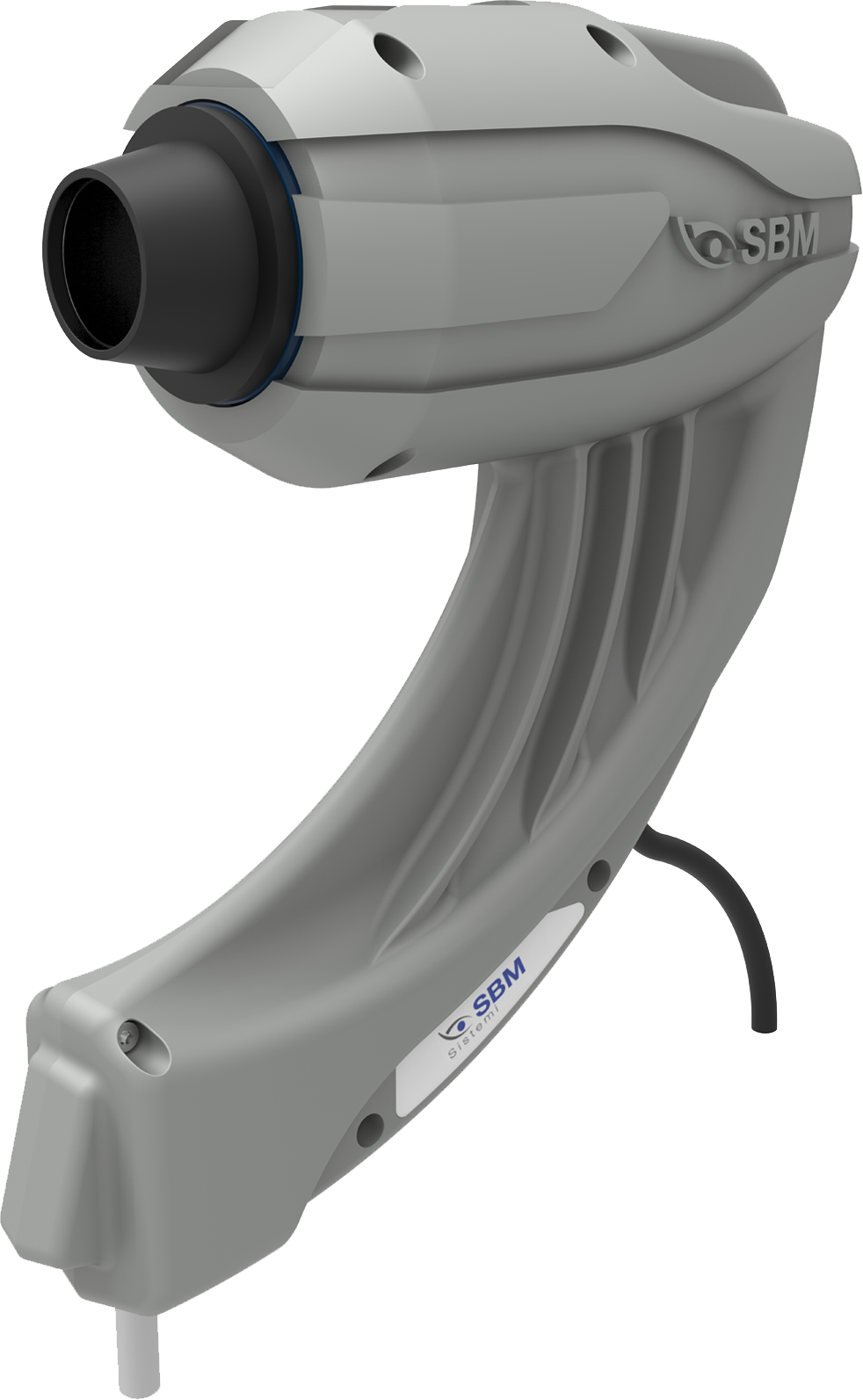
DEM100
- Interferometry Test
- Tear meniscus
- Nibut
- Meibography
- Blepharitis
- Ocular Redness Classification
- Anterior segment imaging
DEM100 PLUS
- Auto Interferometry Test
- Tear meniscus
- Nibut
- Meibography
- 3D Meibography
- Blink Quality
- Blepharitis
- Ocular Redness Classification
- Anterior segment imaging
DSLC only
- Image and video acquisition

OSA Basic
- Interferometry test
- Tear Meniscus
- Auto-NIBUT
- Meibography
- Blepharitis
- Ocular redness classification
- Pupillometry
- White to White measurement
- Anterior segment imaging
OSA Plus
- Interferometry test
- Tear Meniscus
- Auto-NIBUT
- NIBUT map and stability graph
- Meibography
- 3D Meibography
- Blink quality
- Blepharitis
- Ocular redness classification
- Pupillometry
- White to White measurement
- Anterior segment imaging

OSA Vet
- Interferometry test
- Tear Meniscus
- NIBUT
- Meibography
- Blepharitis
- Ocular redness classification
- Pupillometry
- White to White measurement
- Anterior segment imaging

MGD Vet
The images are then automatically classifed.
Thanks to the modular double LED illumination, the image will be taken without interferences.
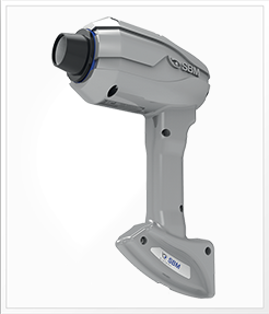
The device is used hand-held and can take pictures of upper and lower lids.

Idra Basic
- Interferometry test
- Tear Meniscus
- Auto-NIBUT
- Meibography
- Blepharitis
- Ocular redness classification
- Pupillometry
- White to White measurement
- Anterior segment imaging
Idra Plus
- Interferometry test
- Tear Meniscus
- Auto-NIBUT
- NIBUT map and stability graph
- Meibography
- 3D Meibography
- Blink quality
- Blepharitis
- Ocular redness classification
- Pupillometry
- White to White measurement
- Anterior segment imaging
Idra Full
- Auto-Interferometry test
- Automatic Tear Meniscus
- Auto-NIBUT
- NIBUT map and stability graph
- Meibography
- 3D Meibography
- Blink quality
- Blepharitis
- Ocular redness classification
- Pupillometry
- White to White measurement
- Anterior segment imaging
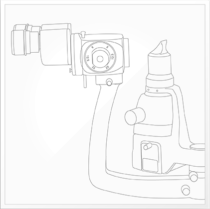
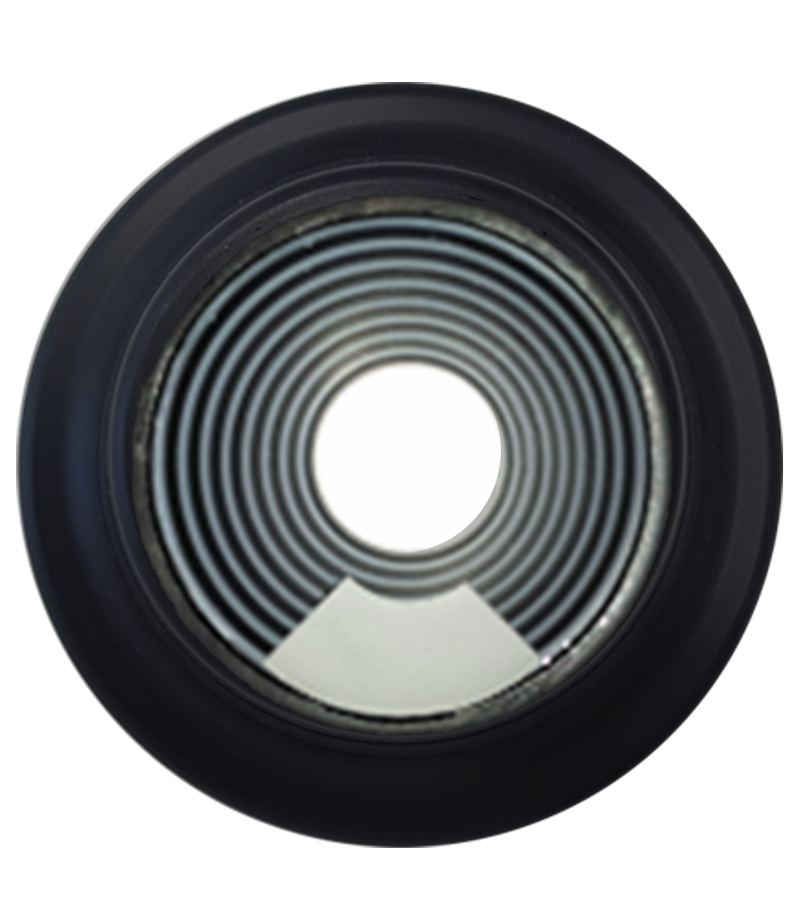
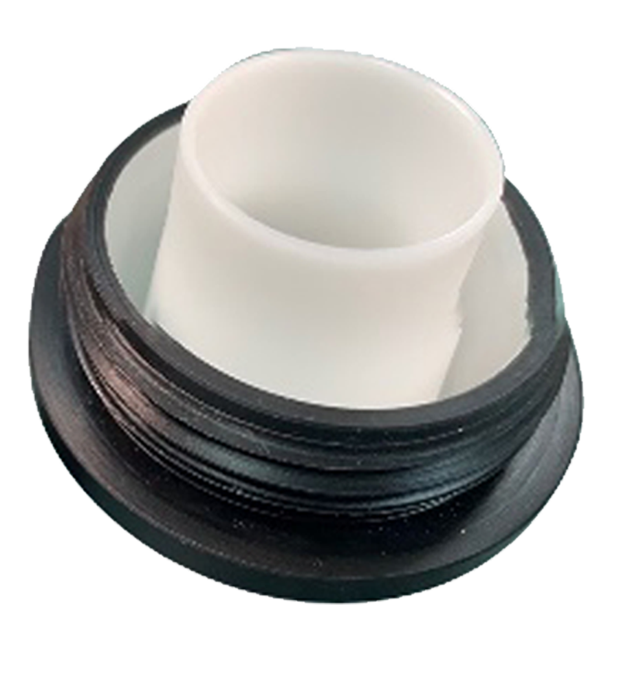
Interferometry cone
Cod.

Placido Disk cone
Cod.
Hybrid cone
Cod. SET/CU
Briefcase
Cod. 313
Dimensions 426x290x159

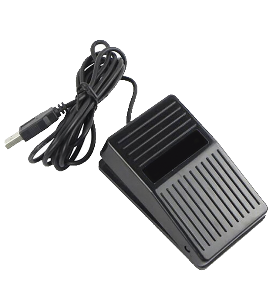
Foot Switch
Cod. 318
To control images and videos acquisition
Interferometry cone
SET/OSAINTCONE
Placido Disk cone
SET/OSAPL

Blepharitis lens
Cod. 322
Magnifying lens to study inflammation conditions of the eyelids

Auto Interferometry cone
SET/333IDRA

Interferometry cone
SET/INTBASE

Placido Disk cone
SET/332IDRAPLACIDO

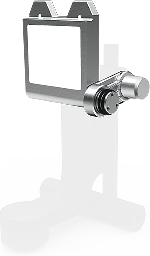
Cones holder
Cod. 338

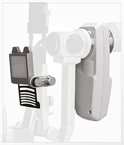
Base and Chin-Rest
Cod. 331+317
To be installed on the ophthalmic table

Base and Chin-Rest
with Baseplate
Cod. 331+344
A complete stand alone support system that requires no

Opthalmic table
Cod. 321
Curved table top (V-shape) with aluminum lifting column, for 2 devices. Table top size 1040mm x 550mm

Holder
Cod. 325
Can be applied on the desktop with two screws to store device when it is not in use

Cones holder
Cod. 338
Blepharitis lens
Cod. 322
Magnifying lens to study inflammation conditions of the eyelids
Foot Switch
Cod. 318
To control images and videos acquisition

Intel® Core™ i7-8565U 4 Core - 1800 MHz 8 GB
DDR 4 (2400 MHz) with 512 GB SSD
Intel® HD Graphics 620
1x HDMI, 1x RJ-45, 1x USB-A 2.0
3x USB-A 3.2 (5 Gbit/s)
2x USB-C 3.2 (5 Gbit/s)
LAN 10/100/1000 Mbit/s
Wi-Fi 5 (802.11ac)
Bluetooth 4.0
Windows 10 PRO
Mini Computer
Cod. PC-0120

of magnification steps
of illumination


